Showing of 1 until 15 from 254 result(s)
Search for: Corneal transplantation; Eye banks; Tissue donors; Organ procurement; Physician's role; Informed consent; Health education; Knowledge, attitudes, practice; Questionnaires
09-fig01.jpg)
Abstract
Objetivo: Analisar o perfil epidemiológico dos casos de evisceração e enucleação no pronto-socorro oftalmológico de um hospital terciário brasileiro.
Métodos: Análise retrospectiva dos casos tratados no pronto-socorro oftalmológico do Hospital São Paulo (Universidade Federal de São Paulo) entre os anos de 2013 a 2018. Os casos urgentes de evisceração e enucleação foram incluídos e os casos eletivos foram excluídos. A análise dos prontuários médicos foi baseada em: dados demográficos, causas imediatas e associadas ao procedimento, acuidade visual informada, duração dos sintomas antes do atendimento oftalmológico, complicações, distância da residência até o hospital e tempo de hospitalização.
Resultados: 61 enucleações e 121 eviscerações foram incluídas no estudo. Os pacientes tinham uma média de idade de 63,27 ± 18,68 anos; 99 eram do sexo masculino (54,50%) e 83 do sexo feminino (45,60%). As indicações de evisceração e enucleação foram: perfuração corneana com (44,50%) e sem (23,63%) sinais infecciosos, endoftalmite (15,38%), trauma ocular (14,29%), neoplasia (0,55%), queimadura (1,10%) e phthisis bulbi (0,55%). A acuidade visual informada foi de ausência de percepção luminosa (87,36%), percepção luminosa (1.10%), ausência de colaboração (3,30%) e sem dados informados (8,24%). A média de tempo até a busca pelo serviço oftalmológico foi de 18,32 dias. Houve 2 casos de oftalmia simpática após evisceração.
Conclusões: Eviscerações foram predominantemente realizadas em comparação a enucleações em todo o período de estudo. As características demográficas mais comuns foram idade >60 anos e sexo masculino. As principais indicações para procedimentos urgentes de evisceração e enucleação foram perfuração corneana com e sem infecção, endoftalmite e trauma ocular. Este estudo poderia guiar medidas preventivas para evitar procedimentos oculares destrutivos.
Keywords: Evisceração do olho; Enucleação ocular; Úlcera da córnea/epidemiologia; Endoftalmite; Traumatismos oculares; Serviços médicos de emergência; Serviços de saúde ocular.
Abstract
PURPOSE: This study aimed to identify the strategies adopted by Brazilian ophthalmologists to control myopia in clinical practice.
METHODS: This was a prospective cross-sectional study. Data were collected using an online questionnaire.
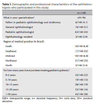
RESULTS: Responses from 148 participants were collected between March and May 2024. The majority of respondents were general ophthalmologists (51%) and pediatric ophthalmologists (43%). They came from all regions of Brazil, but more than half (52%) were from the Southeast region. Most participants (30%) had over 20 years of clinical practice experience. A significant proportion (89.2%) treated progressive myopia. The most requested complementary exams were optical biometry (83.78%) and corneal topography or tomography (69.59%). Behavioral measures were considered the most effective myopia treatment strategies by 41.2% of the respondents, followed by optical (33.8%) and pharmacological interventions (25%). Most recommended spending more time outdoors (94.59%) and reducing screen time (93.92%). Spectacle lenses for myopia (83.11%) and 0.025% atropine eye drops (54.73%) were the most prescribed treatments after the recommendation of environmental and behavioral changes.
CONCLUSION: This study presents a novel analysis of the clinical strategies for myopia control among Brazilian ophthalmologists. Understanding current clinical practices and identifying possible improvements are essential steps toward developing evidence-based guidelines and professional education aimed at improving patient care.
Keywords: Myopia/epidemiology; Refractive errors; Contact lenses; Myopia/drug therapy; Atropine/therapeutic use; Ophthalmologists; Practice patterns, physicians’; Surveys and questionnaires; Brazil/epidemiology
Abstract
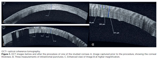
PURPOSE: To assess the reliability and penetration depth of an automated micropuncture system using a tattoo machine.
METHODS: Twenty human corneas were obtained and subjected to intrastromal micropuncture using a tattoo machine. Each cornea was divided into two halves: one received pigment, while the other received saline solution as a control. The Cheyenne tattoo machine was operated at 60 Hz, with standardized needle exposure (six passes per application). The machine used cartridges containing five microneedles. The study was registered with Agência Nacional de Vigilância Sanitária ANVISA (numbers 80281110015, 80281110016, and 80281110019). The pigment used was Electric Ink black ink, with a density of 1,271,460 μg/mL. Puncture depth was measured before and after the procedure using both anterior segment optical coherence tomography and histopathological analysis. Puncture depth measurements were analyzed using ImageJ software. Each cornea was measured thrice, and the results were subsequently compared.
RESULTS: No corneal perforations were observed with the use of the tattoo machine, and puncture depth measurements ranged from 107 to 486 µm.
CONCLUSIONS: The use of a tattoo machine represents a viable and accessible approach for keratopigmentation, with potential for both cosmetic and therapeutic applications. Its adaptation for controlled intrastromal drug delivery may enable the targeted treatment of deep infectious keratitis, corneal neovascularization, and stromal inflammatory disorders, representing a promising approach for corneal stromal diseases. Further research is needed to optimize techniques and evaluate long-term safety and efficacy, particularly for the delivery of antimicrobial, anti-inflammatory, and anti-vascular endothelial growth factor agents.
Keywords: Eye banks; Cadaver; Cornea; Corneal stroma; Drug delivery systems; Tissue donors; Tattooing/instrumentation; Punctures
Abstract
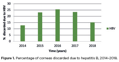
PURPOSE: This study evaluated the proportion of corneas discarded by the Eye Bank of Londrina, Paraná, due to positive serology over a 5-year period and its impact on transplant availability.
METHODS: A cross-sectional study was conducted, analyzing 1,968 corneas from 1,056 donors collected between January 2014 and December 2018 at the Eye Bank of Londrina. Serological tests for hepatitis B (HBsAg and anti-HBc), hepatitis C (anti-HCV), and HIV (anti-HIV 1 and 2) were performed using chemiluminescent microparticle immunoassays. Data were analyzed descriptively and presented in tables and graphs.
RESULTS: Of the 1,968 corneas processed, 897 (45.57%) were discarded. Among these, 333 (37.12%) tested positive for serological markers. Hepatitis B accounted for 34.67% of positive cases (15% of total donations), hepatitis C for 1.11% (0.50% of total), and HIV for 0.89% (0.4% of total). Hepatitis cases remained stable between 2014 and 2016, with a marked decline in 2017 and 2018. Most discarded corneas were positive for anti-HBc (31.88%) and negative for HBsAg; however, the anti-HBs test was not performed to confirm immunity to the hepatitis B virus.
CONCLUSION: The findings highlight the importance of serological testing to identify and eliminate contaminated corneas, thereby preventing the transmission of infectious diseases to recipients. Positive serology for hepatitis, particularly hepatitis B, was the leading cause of corneal disposal.
Keywords: Cornea; Corneal transplantation; Corneal donation; Eye banks; Hepatitis B virus; Hepatitis C virus; HIV infections; Seropositivity; Serologic tests
03-qua01tb.jpg)
Abstract
Objetivos: Dimensionar o impacto da pandemia da COVID-19 nas doações e transplantes de córnea no Brasil e obter indicadores confiáveis para o embasamento de proposições de medidas efetivas para a manutenção e o aperfeiçoamento do sistema de obtenção, processamento, distribuição, utilização e controle dos tecidos oculares doados.
Métodos: Um questionário foi enviado, pelo escritório Brasil da Associação Pan-Americana de Bancos de Olhos (APABO), aos Bancos de Olhos brasileiros. Dados de janeiro a agosto de 2020 foram coletados para gerar indicadores confiáveis sobre o impacto da pandemia da COVID-19 nas doações e transplantes de córnea no Brasil.
Resultados: Dados de 37 Bancos de Olhos mostraram que 76,1% das 3.060 doações e 74,5% dos 3.167 transplantes aconteceram no período pré-pandemia. Das 6.052 córneas processadas 71,8% foram disponibilizadas para fins terapêuticos: 72,9% foram transplantadas, 26,1% acabaram sendo inviabilizadas (45% destas, classificadas para indicações ópticas) e 1%, em glicerina, permanecia em estoque. Das 1.706 córneas que não puderam ser disponibilizadas para uso terapêutico, 47,9% foram excluídas por fatores relacionados às condições dos tecidos, 43,6% por fatores sorológicos, 6,7% por contraindicações constatadas em histórico clínico após a captação e 1,8% por outros fatores.
Conclusões: O impacto negativo da pandemia nas doações e transplantes de córnea no Brasil se deveu à recomendação do Ministério da Saúde de suspender, por quase seis meses, as captações de doadores em parada cardiorrespiratória. Os indicadores tornam evidente a necessidade de atualização dos critérios de classificação e disponibilização das córneas pelos Bancos de Olhos e do sistema nacional de distribuição destes tecidos.
Keywords: Bancos de Olhos; Córnea; Doação de tecidos; Transplante de Córnea; COVID-19; Política pública; Brasil.
14-gra01.jpg)
Abstract
OBJETIVOS: As características físico-químicas e o baixo custo da água de coco foram fundamentais para o este estudo. Analisar o uso de solução a base de água de coco como meio de conservação de córneas humanas em banco de olhos.
MÉTODOS: Estudo experimental e controlado realizado no Banco de Olhos do Hospital Geral de Fortaleza. Utilizou-se solução à base de água de coco preparada no laboratório de Tecnologia de Sêmen de Caprinos do Departamento de Medicina Veterinária da Universidade Estadual do Ceará. Foram usadas córneas de descartes divididas em dois grupos: G1 (Conservante com água de coco) - grupo experimental e G2 (grupo Conservante com OPTISOL GS®) grupo controle, em experimentos sequenciais. A osmolaridade do G1 foi analisada sequencialmente com 275, 300, 325, 345, 365 e 400 mOsm/L. A viabilidade das córneas foram realizadas por microscopia especular e biomicroscopia nos 1º, 3º e 7º dias.
RESULTADOS: As córneas em solução de 365 e 345 mOsm/L apresentavam transparência nos 8mm centrais até o 3º dia, com edema em toda periferia, dobras centrais e edema 2+, com perda parcial do epitélio até 7º dia, sendo o de maior osmolaridade com melhor transparência durante o seguimento. Grupo com 275, 300 e 400 mOsm/L, córnea opaca, edema difuso, perda total do epitélio no 3º dia. As córneas em Optisol mantiveram seus aspectos.
CONCLUSÕES: O conservante à base de água de coco manteve em parte a transparência corneana e a integridade epitelial, especialmente nos primeiros 3 dias de seguimento. A solução conservante com água de coco nas formulações utilizadas não se mostrou eficaz para o uso em banco de olhos humanos.
Keywords: Córnea; Água de coco; Preservação de órgãos/métodos; Solução para preservação de órgãos; Biotecnologia
Abstract
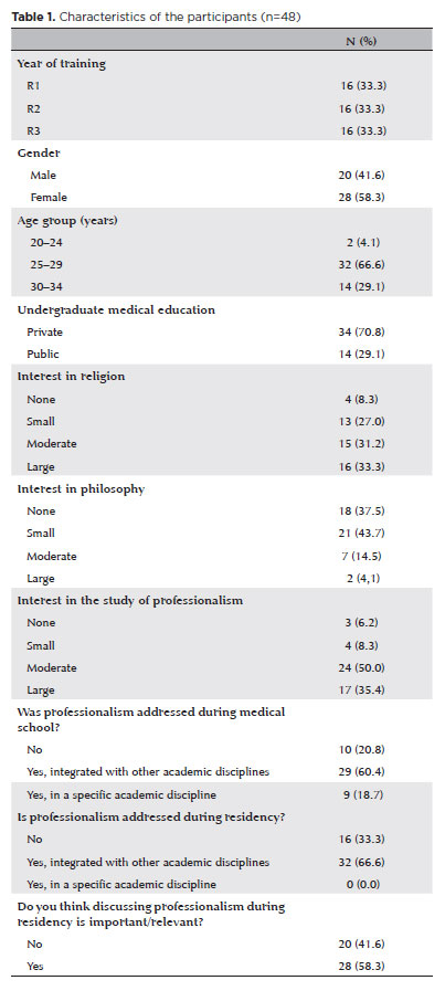
PURPOSE: This study aimed to examine factors related to the professionalism of ophthalmology residents.
METHODS: A cross-sectional study was carried out involving 48 ophthalmology residents in Brazil. Professionalism was assessed using the professionalism mini-evaluation exercise, completed by both preceptors and residents, and the Pennsylvania State College of Medicine Professionalism Questionnaire, completed by the residents. The association between the professionalism score assigned by the preceptor through the professionalism mini-evaluation exercise and various sociodemographic and educational variables was assessed. The correlation between the residents’ self-assessment across both instruments and the preceptor’s assessments was measured using Spearman’s Rho.
RESULTS: All 48 residents were included, with equal representation across the 3 years of residency. The majority were female (58.3%) and between 25 and 29 years old (66.7%). The average professionalism score on the professionalism mini-evaluation exercise given by the preceptors was 3.0 (75%). A significant association was found between the year of training and the score in the doctor-patient relationship domain, with first-year residents showing lower scores (p=0.002). Male residents had higher scores in the “Interprofessional” domain (p=0.031). Graduates from private medical schools scored higher in both the “doctor-patient relationship” (p=0.015) and “reflective skills” (p=0.033) domains. Lower interest in professionalism was linked to lower scores in the “Interprofessional relationships” (p=0.033) and “time management” (p=0.003) domains. A strong correlation was observed between preceptor’s professionalism mini-evaluation exercise scores and residents’ self-assessed professionalism mini-evaluation exercise scores (r=0.917). However, the correlation between the self-assessed professionalism mini-evaluation exercise and the Pennsylvania questionnaire scores was weak (r=0.226).
CONCLUSION: Professionalism scores among ophthalmology residents were associated with year of training, gender, type of undergraduate education, and level of interest in the topic.
Keywords: Internship and residency; Ophthalmology; Professional competence; Education, professional; Physician-patient relations; Surveys and questionnaires
Abstract
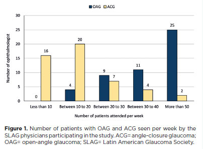
PURPOSE: To evaluate the preferred surgical practice patterns for glaucoma among members of the Latin American Glaucoma Society.
METHODS: A cross-sectional study was conducted using an electronic survey distributed in July 2023 via email to members of the Latin American Glaucoma Society. The questionnaire comprised four sections addressing the specialists' profiles, preferred surgical procedures for open-angle glaucoma, and choices in 10 different clinical scenarios, including congenital glaucoma.
RESULTS: Of the 63 members, 49 physicians (77.7%) responded – 13 women and 36 men – from nine Latin American countries. Thirty-one respondents (63.26%) had more than 20 yr of professional experience. For the surgical management of open-angle glaucoma, trabeculectomy was the most preferred procedure (48 physicians), followed closely by glaucoma drainage devices (47 physicians) and minimally invasive glaucoma surgery (29 physicians). Across the 10 clinical scenarios, glaucoma drainage devices were selected most frequently (203 preferences), followed by trabeculectomy (118), ciliary body laser procedures (107), and minimally invasive glaucoma surgery (40). However, minimally invasive glaucoma surgery was the preferred option for primary open-angle glaucoma with mild-to-moderate cataracts.
CONCLUSION: Among specialists of the Latin American Glaucoma Society, trabeculectomy and glaucoma drainage devices remain the most commonly performed surgical procedures. Minimally invasive glaucoma surgery is primarily used in combination with cataract surgery, while ciliary body laser procedures are generally reserved for cases of previous glaucoma drainage device failure or as an initial option for newly diagnosed glaucoma cases.
Keywords: Glaucoma; Ophthalmologic surgical procedures; Latin America; Practice patterns, physicians; Surveys and questionnaires
Abstract
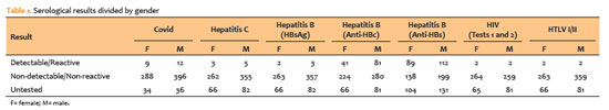
PURPOSE: To evaluate the impact of the COVID-19 pandemic and characterize the serological profile of discarded corneal donations in the coverage area of the Banco de Olhos de Londrina, through reverse transcription-polymerase chain reaction testing for COVID-19 and serological screening of cornea donors excluded because of positive test results.
METHODS: This observational retrospective study included 776 cornea donors who’s serological and reverse transcription-polymerase chain reaction test results were processed at the Hospital of Universidade Estadual de Londrina between May 2020 and 2022. The number of corneal donations and tissue utilization rates throughout the years of operation of the Banco de Olhos de Londrina were also analyzed.
RESULTS: The mean donor age was 53.14 years; 332 donors (43%) were female, and 444 (57%) were male. Positive results were identified in 15.76% of donors for hepatitis B core antibody antibodies, 0.65% for hepatitis B surface antigen, 1.03% for hepatitis C antibodies, and 0.52% for human immunodeficiency virus and human T-lymphotropic vírus. Positive reverse transcription-polymerase chain reaction results for SARS-CoV-2 were observed in 2.7% of cases. Older adults were 2.6 times more likely to test positive for SARS-CoV-2 (95% CI, 1.06-6.34) and 3.0 times more likely to test positive for hepatitis B core antibody (95% CI, 1.95-4.41) than younger individuals. A 75.2% reduction in corneal donations was observed in 2020 compared with 2019, accompanied by a 5% increase in tissue utilization, possibly associated with the effectiveness of donor screening during the pandemic.
CONCLUSION: The COVID-19 pandemic had a profound impact on the number of corneal transplants worldwide, in Brazil, and at the Banco de Olhos de Londrina because of the substantial decline in donations during this period. Hepatitis B was the leading cause of corneal tissue discard due to positive serology in both this study and previous reports, highlighting the importance of prevention programs and improved vaccination coverage. Strict legislation, comprehensive serological screening, and appropriate processing of donated tissue remain essential to eliminate potential sources of infection and ensure transplantation safety.
Keywords: Cornea; Corneal transplantation; COVID-19; Eye banks; Serology
Abstract
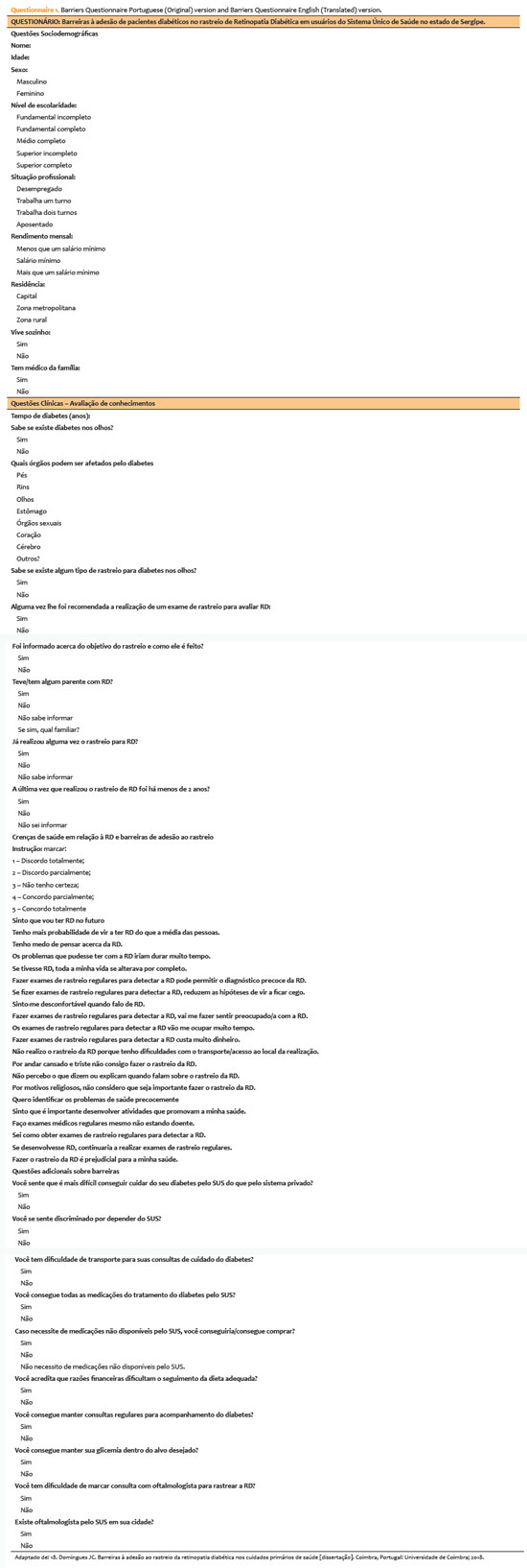
PURPOSE: This study aimed to identify barriers to diabetic retinopathy screening among a socioeconomically vulnerable urban population in northeast Brazil.
METHODS: A cross-sectional study was conducted during a diabetic retinopathy screening campaign at primary healthcare units. Ninety-five patients with diabetes underwent retinal examinations and completed a structured interview. Clinical, demographic, and socioeconomic data were collected.
RESULTS: The study population consisted predominantly of older adults (mean age: 60.7 ± 10.5 years), with a high prevalence of type 2 diabetes (99.0%) and low educational attainment. Most participants were economically inactive (81.1%) and reported low income (83.2%). Diabetic retinopathy and maculopathy were highly prevalent, affecting 50.0% and 22.9% of participants, respectively. Longer duration of diabetes was significantly associated with greater awareness of diabetic retinopathy (p=0.035), higher HbA1c levels (p<0.001), and increased prevalence of diabetic retinopathy (p=0.013) and maculopathy (p=0.002). Notably, 33.3% of participants reported difficulties attending medical appointments for diabetes management. In addition, 78.1% experienced challenges scheduling ophthalmologic evaluations, and 76.3% reported that no ophthalmologist was available in their city through the public healthcare system. Financial constraints also limited adherence to recommended dietary practices (90.4%) and impaired glycemic control, with more than half of participants reporting difficulty maintaining target glucose levels.
CONCLUSION: Major barriers to diabetic retinopathy screening included limited awareness of the importance of screening, financial hardship, and transportation challenges. Targeted educational initiatives and structural interventions such as expanded screening programs incorporating telemedicine and subsidized transportation—may improve screening adherence among vulnerable populations.
Keywords: Diabetic retinopathy; Mass screening; Health services accessibility; Health knowledge, attitudes, practices; Socioeconomic factors
14-tab01tb.jpg)
Abstract
OBJETIVOS: À medida que a utilização de equipamentos digitais no emprego aumenta, a avaliação do seu efeito na saúde visual necessita de ferramentas válidas e robustas. Este estudo teve como objetivo traduzir, adaptar culturalmente e validar para português o Questionário da Síndrome Visual do Computador (CVS-Q©).
MÉTODOS: O procedimento foi realizado em 5 fases: tradução direta, síntese da tradução, tradução inversa, consolidação por um painel de especialistas, e pré-teste. Para fazer o pré-teste foi realizado um estudo piloto transversal aplicado a uma amostra de 26 participantes que completaram a versão pré-final da versão portuguesa do CVS-Q©, questionando por dificuldades, compreensão e sugestões de melhoria do questionário. Para avaliar a confiança e validade da versão portuguesa do CVS-Q©foi realizado um estudo transversal de validação em uma amostra diferente (280 funcionários).
RESULTADOS: No pré-teste, 96.2% dos participantes não apresentaram dificuldades no preenchimento do questionário, enquanto 84.0% indicaram que era claro e compreensível. Obteve-se, então, o CVS-Q© em português (Questionário da Síndrome Visual do Computador, CVS-Q PT©). A sua validação revelou uma boa consistência interna
da sua escala (Cronbach's alpha=0.793), boa estabilidade temporal (coeficiente de correlação interclasse=0.847; 95% CI 0.764-0.902, kappa=0.839), sensibilidades e especificidades adequadas (78.5% e 70.7%, respetivamente), boa capacidade de discriminação (área abaixo da curva=0.832; 95% CI 0.784-0.879), e uma adequada validade da convergência com o índice de doença da superfície ocular (ocular surface disease index - OSDI; coeficiente de correlação de Spearman=0.728, p<0.001). A análise fatorial revelou um único fator responsável por explicar a variância comum em 37.7%. Um funcionário com uma pontuação ≥7 pontos sofria de síndrome visual do computador.
CONCLUSÃO: O CVS-Q PT© pode ser considerada uma ferramenta intuitiva, de fácil interpretação e com boas propriedades psicométricas para avaliar a síndrome visual do computador em funcionários portugueses expostos a ecrãs digitais. Este questionário facilitará as decisões sobre medidas preventivas, intervenções e tratamento, e a comparação entre as populações expostas em diferentes países de língua portuguesa.
Keywords: Síndrome visual do computador; Dispositivos digitais; Saúde ocular; Estudo de validação; Propriedades psicométricas; Inquéritos e questionários
Abstract
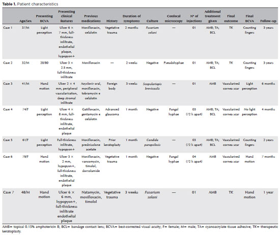
PURPOSE: This study aimed to report the use, efficacy, and safety of intracameral voriconazole as an adjuvant treatment for deep fungal keratitis.
METHODS: This was a prospective case series of seven eyes with fungal keratitis with anterior chamber involvement or a corneal ulcer refractory to conventional topical treatment. In addition to topical treatment with 0.15% amphotericin B eye drops, voriconazole 50 μg/ 0.1 mL
was administered to the anterior chamber of each affected eye up to four times within 72 h. The primary outcome measures were healing (fungal eradication) and the need for therapeutic keratoplasty. Best-corrected visual acuity was a secondary outcome measure.
RESULTS: Three cases were confirmed by confocal microscopy, and four were diagnosed from positive culture tests. At presentation, one patient had a best-corrected visual acuity of 20/80, while all others had hand motion or worse. Four cases received one intracameral injection, two cases received three, and one case received four injections. There were no complications after any of the intracameral voriconazole injections. Four patients had imminent corneal perforations and were treated with cyanoacrylate adhesive and bandage contact lenses. Four patients recovered from the infection, and three underwent therapeutic keratoplasty. The final best-corrected visual acuity was improved in two cases but all patients had a final visual acuity of counting fingers or worse.
CONCLUSION: As an adjuvant treatment for deep fungal keratitis, intracameral voriconazole injection is a feasible option. Although fungal eradication was achieved in all patients, three required therapeutic keratoplasty and all patients had unsatisfactory visual acuity outcomes.
Keywords: Antifungal agents; Fungi; Corneal transplantation; Keratitis; Eye infections, fungal; Voriconazole
Abstract
PURPOSE: To evaluate the clinical results of cryopreserved amniotic membrane transplantation as a treatment option for refractory neurotrophic corneal ulcers.
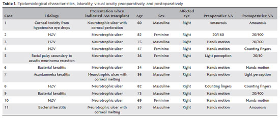
METHODS: This prospective study included 11 eyes of 11 patients who underwent amniotic membrane transplantation for the treatment of refractory neurotrophic corneal ulcers at Hospital de Clínicas da Universidade Federal do Paraná, in the city of Curitiba, from May 2015 to July 2021. Patients underwent different surgical techniques in which the amniotic membrane was applied with the epithelium facing upward to promote corneal re-epithelialization.
RESULTS: The median age of the patients was 60 years (range, 34-82 years), and 64% were men. The predominant etiology of corneal ulcers was herpes zoster (45% of cases). Approximately one-third of the patients (27%) were chronically using hypotensive eye drops, and more than half (54%) had previously undergone penetrating corneal transplantation. At the time of amniotic membrane transplantation, 18% of the eyes had corneal melting, 9% had corneal perforation, and the others had corneal ulceration without other associated complications (73%). The time between clinical diagnosis and surgical treatment ranged from 9 days to 2 years. The corrected visual acuity was worse than 20/400 in 90% of the patients preoperatively, with improvement in 36% after 3 months of the procedure, worsening in 18% and remaining stable in 36%. Of the patients, 81% complained of preoperative pain, and 66% of them reported total symptom relief after the surgical procedure. In one month, 54.6% of the patients presented a closure of epithelial defect, and half of the total group evolved with corneal thinning. The failure rate was 45.5% of the cases.
CONCLUSION: Cryopreserved amniotic membrane transplantation can be considered a good alternative for treating refractory neurotrophic corneal ulcers, as it resulted in significant improvement in pain (66%) and complete epithelial closure (60%) in many patients at 1 month postoperatively. Notably, the high failure rate highlights the need for further studies to identify patient- and ulcer-related factors that may influence the outcomes of this procedure.
Keywords: Amnion/transplantation; Corneal ulcer; Anterior eye segment; Keratitis
14-fig01.jpg)
Abstract
O presente trabalho traz uma revisão das abordagens terapêuticas para a cegueira da córnea. O estudo detalha as etapas e os elementos envolvidos na cicatrização da córnea. Ele mostra as limitações das estratégias cirúrgicas e farmacológicas usadas para restaurar e manter a transparência da córnea em termos de sobrevida a longo prazo e alcance geográfico. As perspectivas dos agentes anabólicos, incluindo vitamina A, hormônios, fatores de crescimento e novos moduladores pró-mitóticos e anti-inflamatórios para auxiliar a cicatrização da ferida na córnea, são revisadas criticamente. Aqui, apresentamos estudos envolvendo nanotecnologia, terapia gênica e reengenharia de tecidos como possíveis estratégias futuras para atuar de maneira isolada ou combinada com a cirurgia da córnea para prevenir ou reverter a cegueira corneana.
Keywords: Cegueira; Doenças da córnea; Transplante de córnea; Terapia genética; Terapia baseada em transplante de células e tecidos; Células-tronco
Abstract
Os bancos de olhos utilizam procedimentos estéreis na manipulação dos olhos, medidas antissépticas para a descontaminação da superfície ocular e critério rigoroso de seleção do doador. Essa seleção é feita por meio do prontuário médico e de testes sorológicos específicos post mortem. Para orientá-la e uniformizá-la, as associações de bancos de olhos e órgãos governamentais fornecem listas de contraindicações absolutas e relativas de uso do tecido, baseadas nas condições prévias de saúde do doador. Essas listas são as guardiãs do princípio de Hipócrates "primum non nocere" e, como tal, são conservadoras. Entretanto, cada transplante traz o risco de transmissão de agentes potencialmente nocivos ao receptor. O objetivo não é eliminar esse risco, mas limitá-lo a um nível razoável. Existe um equilíbrio entre a segurança e a disponibilidade de córneas. A sabedoria está em manter esse equilíbrio, exercendo a prudência sem rigor exagerado.
Keywords: Bancos de olhos/normas; Transplante de córnea; Seleção do doador; Coleta de tecidos e órgãos
 ABO is licensed under a Creative Commons Attribution-NonComercial 4.0 Internacional.
About
Issues
Editorial Board
Submission
Arquivos Brasileiros de Oftalmologia
Official publication of Brazilian Council of Ophthalmology - Conselho Brasileiro de Oftalmologia (CBO)
Rua Casa do Ator, 1.117 - 2nd floor - Zip Code: 04546-004
São Paulo - SP, Brazil
TEL: +55 11 3266-4000
E-mail: [email protected]