Arq. Bras. Oftalmol. 2023;86 (4 )
:308-313
| DOI: 10.5935/0004-2749.20230052
Abstract
Objetivo: Apresentar os resultados de um estudo retrospectivo sobre o impacto clínico e econômico da administração de cefuroxima intracameral para prevenir endoftalmite nas cirurgias de catarata em um hospital de referência.
Métodos: Este estudo incluiu 16.902 olhos de pacientes submetidos à cirurgia de catarata entre 2013 e 2017. A partir de maio de 2014, todos os pacientes receberam rotineiramente uma injeção intracameral de 1mg de cefuroxima (10mg/1mL) ao final da cirurgia de facoemulsificação. A eficácia da profilaxia foi avaliada usando o risco relativo e o impacto econômico foi avaliado com o número necessário para tratar para se evitar um caso de endoftalmite.
Resultados: Antes da introdução do protocolo da cefuroxima, foram realizadas 3.407 cirurgias de catarata por facoemulsificação e ocorreram 7 casos de endoftalmite pós-operatória (incidência de 0,2%). Após a introdução do protocolo da cefuroxima, foram realizadas 13.495 cirurgias e registrados 4 casos de endoftalmite (incidência de 0,03%). A cefuroxima foi um fator de proteção no desenvolvimento de endoftalmite (risco relativo = 14%, p=0,002, Intervalo de Confiança de 95% [IC 95%], 4% - 49%) e o impacto econômico do número necessário para tratar = 568. A economia potencial com a cefuroxima foi de aproximadamente US$ 2.334,36 para cada 568 pacientes tratados.
Conclusão: A incidência de endoftalmite diminuiu 86% (risco relativo = 14%, p=0,002, IC 95% 4% - 49%) desde a introdução da profilaxia com cefuroxima intracameral no hospital do estudo. Os resultados apresentados mostram forte evidência para o uso da cefuroxima na profilaxia da endoftalmite após cirurgias de facoemulsificação, por proporcionar economia de custos e benefício clínico.
Keywords: Extração de catarata; Endofitalmite; Cefuroxima; Antibioticoprofilaxia; Avaliação de custo-efetividade.
Arq. Bras. Oftalmol. 2024;87 (1 )
:1-6
| DOI: 10.5935/0004-2749.2021-0311
Abstract
Objetivo: Esta pesquisa buscou determinar o impacto dos níveis de proteína G sérica no desenvolvimento da retinopatia em pacientes diabéticos, comparando-os a indivíduos saudáveis.
Métodos: Foram incluídos, no estudo, 40 pacientes com retinopatia diabética (Grupo 1), 40 pacientes sem retinopatia diabética (Grupo 2) e 40 indivíduos saudáveis (Grupo 3). Os níveis hormonais de progesterona sérica, de proteína G sérica, estradiol, oxidante/antioxidante e hormônio liberado pela tireoide foram analisados e comparados. A análise post hoc foi realizada para comparar os subgrupos nos quais diferenças estatisticamente significativas foram encontradas.
Resultados: Uma diferença significativa foi encontrada entre todos os grupos em termos de proteína G sérica, oxidante/antioxidante e níveis de estradiol (p<0.01), mas nenhuma diferença significativa foi encontrada em termos de hormônio liberado pela tireoide ou progesterona (p=0,496, p=0,220, respectivamente). Na análise post hoc dos grupos com diferenças estatisticamente significativas, outra diferença significativa foi encontrada entre todos os grupos para proteína G sérica e níveis oxidantes/antioxidantes (p<0,05). Os níveis de proteína G sérica e os níveis de oxidante foram positivamente correlacionados, enquanto os níveis de proteína G sérica e os níveis de antioxidantes foram negativamente correlacionados (r=0,622/p<0,01, r=0,453/p<0,01, r=0,460/p<0,01, respectivamente). A análise de regressão múltipla mostrou que o aumento da proteína G sérica pode ajudar a prevenir a retinopatia diabética.
Conclusões: Os níveis de proteína G sérica que eram mais altos no grupo de retinopatia diabética, aumentaram à medida que o equilíbrio oxidante/antioxidante mudou em favor do estresse oxidativo. Este parece ser um mecanismo de defesa para prevenir danos neuronais.
Keywords: Retinopatia diabética; GPER-1; Estradiol; Progesterona; Estresse oxidativo; Oxidantes.
Arq. Bras. Oftalmol. 2022;85 (2 )
:136-143
| DOI: 10.5935/0004-2749.20220022
Abstract
Objetivo: Estimar a epidemiologia do pterígio; sua correlação com sintomas de olho seco e com potenciais preditores sistêmicos e oculares.
Métodos: Estudo transversal, de base populacional, no qual foram realizadas visitas domiciliares aleatórias a 600 participantes, com 40 anos ou mais de idade, em Ribeirão Preto-SP (n=420) e Cassia dos Coqueiros-SP (n=180), Brasil. Uma entrevista estruturada com um questionário detalhado foi usada para coletar informações sobre demografia e possíveis fatores de risco. Em um segundo momento, participantes aleatórios com pterígio (n=63) ou não (n=110) foram avaliados quanto a alterações na superfície ocular.
Resultados: A frequência de pterígio em Ribeirão Preto foi de 21%; 15.7% entre as mulheres e 32.1% entre os homens (p=0,0002). Em Cássia dos Coqueiros, essa taxa foi de 19.4%; onde 17.3% eram mulheres e 25.5% eram homens (p=0,28). A média de idade naqueles afetados pelo pterígio foi superior à dos participantes sem pterígio, 65,6 ± 10,5 e 61,2 ± 12,0 anos, respectivamente (p=0,02). Houve uma correlação positiva entre o pterígio e história prévia de radioterapia e quimioterapia (p<0,0001 para ambos). Houve maior coloração de fluoresceína na córnea e maior coloração de lissamina verde na conjuntiva em olhos com pterígio (p=0,0003 e 0,0001, respectivamente).
Conclusão: Encontramos uma alta frequência de pterígio em duas populações adultas brasileiras, principalmente em homens e idosos. Danos na superfície ocular e história prévia de radioterapia e/ou quimioterapia foram associados ao pterígio.
Keywords: Pterígio/epidemiologia; Síndrome do olho seco; Prevalência; Fatores de risco
Arq. Bras. Oftalmol. 2022;85 (4 )
:389-398
| DOI: 10.5935/0004-2749.20220042
Abstract
Objetivos: Explorar sistematicamente as mudanças dinâmicas e a sequência temporal no processo de apoptose de células epiteliais corneanas após excesso de irradiação com ultravioleta B.
Métodos: A radiação ultravioleta B (144 mJ/cm2) foi utilizada para irradiar células epiteliais da córnea de rato durante 2h. A morfologia celular foi observada por meio de microscópio de contraste de interferência diferencial, e os números de diferentes tipos de células apoptóticas foram contados e registrados pelo software ImageJ. A viabilidade celular foi medida pelo método brometo de 3- (4, 5-dimetil-2-tiazolil) -2, 5-difenil-2-H-tetrazólio. A taxa apoptótica celular e a perda do potencial da membrana mitocondrial foram detectadas por meio de análises citométricas de fluxo. Os níveis de expressão de três genes apoptóticos foram medidos por reação em cadeia da polimerase quantitativa em tempo real em diferentes momentos dentro de 0-24 h após a irradiação.
Resultados: Após 144 mJ/cm2 de irradiação com ultravioleta B por 2h, os níveis de expressão de caspase-8 e Bax foram maiores em 0h; o potencial da membrana mitocondrial diminuiu a 0h e permaneceu constante por 6h na cultura subsequente. Às 6h, a caspase-3 foi ativada. A diminuição da viabilidade celular e o aumento da taxa apoptótica atingiu o pico em 6h. A expressão de caspase-3 diminuiu dentro de 12 - 24 h, levando a um declínio na taxa apoptótica e alteração no estágio apoptótico.
Conclusões: As células epiteliais da córnea apresentaram uma apoptose rápida após excesso de irradiação com ultravioleta B, e esse processo foi associado tanto à via extrínseca como à via intrínseca.
Keywords: Irradiação com ultravioleta B; Radiação; Epitélio anterior; Célula epitelial; Sobrevivência celular; Apoptose; Ratos
Arq. Bras. Oftalmol. 2026;89 (2 )
:1-8
| DOI: 10.5935/0004-2749.2025-0175
Abstract
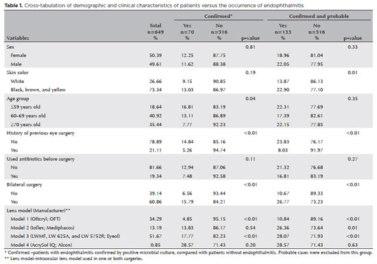
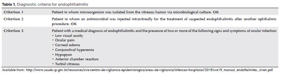
PURPOSE: Endophthalmitis is one of the most important adverse events after cataract surgery, as it can lead to total vision loss. This study aimed to describe the occurrence of endophthalmitis after phacoemulsification with intraocular lens implantation in patients treated in a community setting in Porto Velho, Rondônia, Brazil.
METHODS: This retrospective cohort study was conducted using a database of 649 medical records of patients who underwent surgery and were followed for three months. Poisson regression analysis was used to estimate relative risks and 95% confidence intervals (95% CIs).
RESULTS: The incidence of confirmed endophthalmitis was 11.94% (95% CI, 9.50-14.76), while the incidence of confirmed and probable cases was 20.50% (95% CI, 17.52-23.73). For confirmed cases, bilateral surgery and the use of lens model 3 were identified as risk factors for endophthalmitis, whereas age over 70 yr and preoperative antibiotic use were protective factors. For confirmed and probable cases, brown and yellow skin color, bilateral surgery, and the use of lens model 3 were also identified as risk factors. Gram-negative bacteria were the predominant etiological agents, and corneal edema was the main clinical manifestation. The mean duration of treatment was eight days, and 27.12% of patients used antibiotics.
CONCLUSION: The incidence observed was substantially higher than that reported in the literature, with a predominance of Gram-negative agents and an association with bilateral surgeries and the Eyeol intraocular lens model. These findings reinforce the need for continuous epidemiological surveillance and the implementation of specific biosafety and infection control protocols during cataract surgery campaigns.
Keywords: Endophthalmitis; Disease outbreaks; Phacoemulsification; Lens implantation, intraocular; Lenses, intraocular; Cataract; Risk factors; Anti-bacterial agents
Arq. Bras. Oftalmol. 2025;88 (6 )
:1-7
| DOI: 10.5935/0004-2749.2025-0083
Abstract
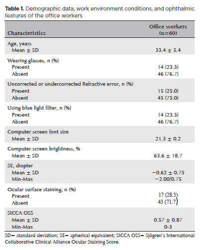
PURPOSE: To examine how ophthalmological features, screen exposure duration, and break habits among office employees affect ocular surface parameters.
METHODS: This single-center cross-sectional study involved two assessments on the same day: one before and one after a visual display terminal task. During the initial assessment, information on screen use was gathered, and refractive error, anterior segment examination, tear breakup time, and Schirmer test measurements were conducted. Participants tracked their screen usage and break durations throughout the day. At the end of the workday, tear breakup time and Schirmer I tests were repeated. Baseline and follow-up results were compared, and regression analysis was performed to identify factors linked to tear breakup time reduction.
RESULTS: The study enrolled 60 female office employees. Their mean screen time was 269.26 ± 70.21 min, with an average break duration of 151.93 ± 46.24 min. Tear breakup time at the second assessment (6.38 ± 2.70) was significantly lower than at baseline (8.62 ± 2.73) (p<0.001), whereas Schirmer test scores showed no significant change (p>0.05). Tear breakup time reduction was noted in 54 participants (90.0%), with a significant association between tear breakup time decrease percentage and screen exposure (p=0.001, r=0.463). Regression analysis showed that uncorrected or undercorrected refractive error was an independent risk factor for a ≥30% tear breakup time reduction, while taking more frequent short breaks (<15 min) acted as a protective factor.
CONCLUSIONS: Taking more frequent short breaks (<15 min) and correcting refractive errors help prevent intra-day tear breakup time decline during visual display terminal use. Structuring breaks to support tear film stability is advisable for occupations that require regular visual display terminal tasks.
Keywords: Tear film; Screen time; Tear breakup time; Office workers; Protective factors; Lacerations; Refractive errors; Risk factors.
Arq. Bras. Oftalmol. 2025;88 (2 )
:1-7
| DOI: 10.5935/0004-2749.2023-0265
Abstract
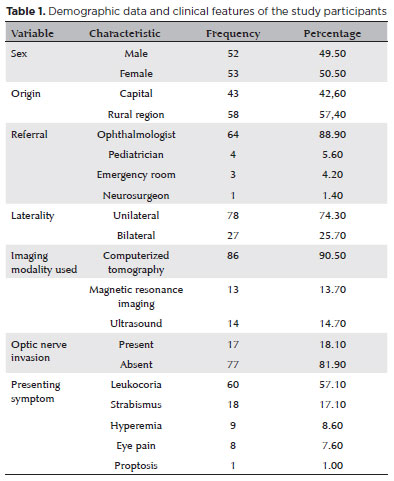
PURPOSE: Although Brazil has a high prevalence of retinoblastoma, there is a lack of epidemiological data on the disease. Thus, in this study, we aimed to evaluate the epidemiological profile of patients diagnosed with retinoblastoma in the ophthalmology department of a pediatric tertiary referral hospital in Ceara, Brazil.
METHODS: A descriptive and cross-sectional study was conducted by retrospectively analyzing the clinical and socioeconomic data from the medical records of pediatric patients followed-up at the hospital between 2007 and 2021. Retinoblastoma was diagnosed on the basis of a fundoscopic or histopathologic examination.
RESULTS: The data of 105 patients were included in the study, and the mean patient age at the time of diagnosis was 1.7 years. Most of the patients were women (50.5%) and hailed from rural areas (57.4%), which was associated with a higher tumor stage. Of the 150 patients, 57.1% initially presented with leukocoria. Ocular hyperemia was associated with more advanced stages of retinoblastoma (p=0.004). Bilateral involvement was observed in 25.7% of the patients and at a significantly younger age (p=0.009). The presence of retinal detachment, vascularized lesions, and vitreous seeds significantly increased the likelihood of requiring enucleation.
DISCUSSION: This study presents an epidemiological description of retinoblastoma in Brazil, which highlights the significance of early detection. Delayed diagnosis is associated with a poorer visual prognosis and higher mortality rate, particularly in patients with unilateral disease. Risk factors for a more severe disease were retinal detachment, vascularized lesions, and vitreous seeds. The correlation between histopathological features and clinical outcomes was limited.
CONCLUSION: Further studies are required to assess the influence of ocular hyperemia, fundoscopic assessment, and histopathologic findings on the prognosis of retinoblastoma. Moreover, it is critical to devise interventions to reduce the time-to-diagnosis in rural areas.
Keywords: Retinoblastoma; Retinal neoplasms; Epidemiology; Prevalence; Risk factors; Delayed diagnosis; Child
Arq. Bras. Oftalmol. 2026;89 (3 )
:1-14
| DOI: 10.5935/0004-2749.2025-0248
Abstract
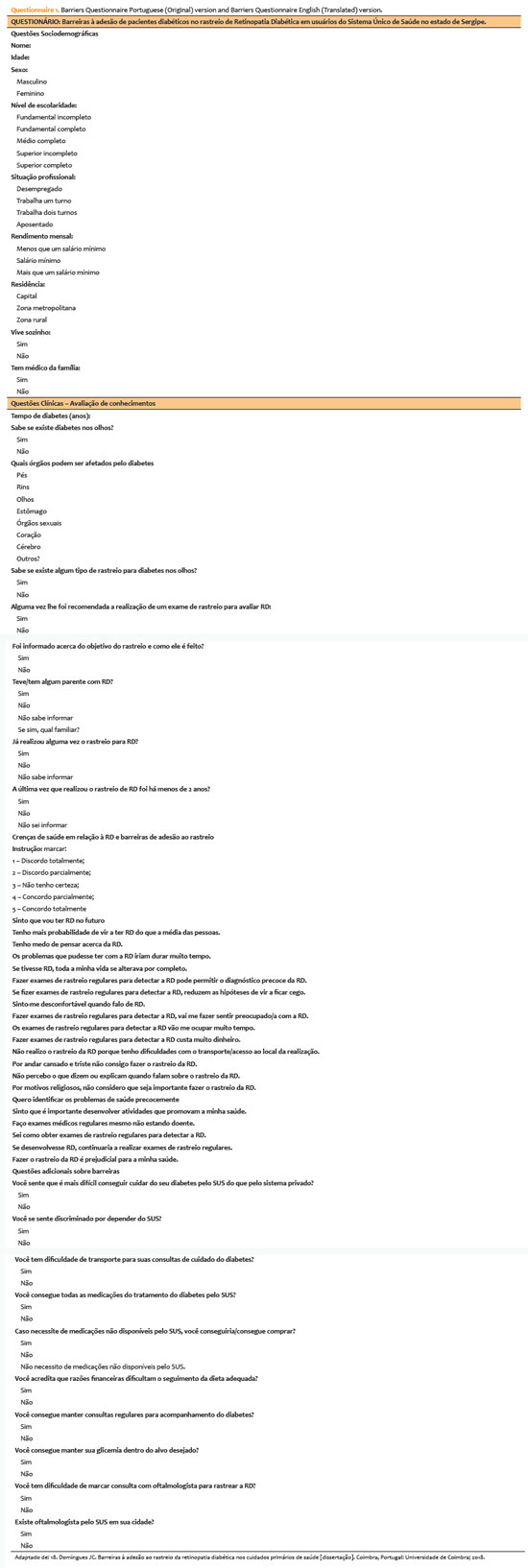
PURPOSE: This study aimed to identify barriers to diabetic retinopathy screening among a socioeconomically vulnerable urban population in northeast Brazil.
METHODS: A cross-sectional study was conducted during a diabetic retinopathy screening campaign at primary healthcare units. Ninety-five patients with diabetes underwent retinal examinations and completed a structured interview. Clinical, demographic, and socioeconomic data were collected.
RESULTS: The study population consisted predominantly of older adults (mean age: 60.7 ± 10.5 years), with a high prevalence of type 2 diabetes (99.0%) and low educational attainment. Most participants were economically inactive (81.1%) and reported low income (83.2%). Diabetic retinopathy and maculopathy were highly prevalent, affecting 50.0% and 22.9% of participants, respectively. Longer duration of diabetes was significantly associated with greater awareness of diabetic retinopathy (p=0.035), higher HbA1c levels (p<0.001), and increased prevalence of diabetic retinopathy (p=0.013) and maculopathy (p=0.002). Notably, 33.3% of participants reported difficulties attending medical appointments for diabetes management. In addition, 78.1% experienced challenges scheduling ophthalmologic evaluations, and 76.3% reported that no ophthalmologist was available in their city through the public healthcare system. Financial constraints also limited adherence to recommended dietary practices (90.4%) and impaired glycemic control, with more than half of participants reporting difficulty maintaining target glucose levels.
CONCLUSION: Major barriers to diabetic retinopathy screening included limited awareness of the importance of screening, financial hardship, and transportation challenges. Targeted educational initiatives and structural interventions such as expanded screening programs incorporating telemedicine and subsidized transportation—may improve screening adherence among vulnerable populations.
Keywords: Diabetic retinopathy; Mass screening; Health services accessibility; Health knowledge, attitudes, practices; Socioeconomic factors
Arq. Bras. Oftalmol. 2025;88 (3 )
:1-6
| DOI: 10.5935/0004-2749.2023-0345
Abstract
PURPOSE: To determine the impact of prophylactic intracameral cefuroxime administration on the post-cataract surgery endophthalmitis rates and analyze its safety.
METHODS: The incidence of post-phacoemulsification endophthalmitis before and after the introduction of antibiotic prophylaxis with cefuroxime was compared. Data were extracted from the electronic medical records of patients who underwent cataract surgery between July 2019 and July 2022 at a tertiary-care hospital. Data were also collected from the Hospital Infection Control Service database. Statistical analysis was performed to assess the efficacy of cefuroxime prophylaxis in reducing endophthalmitis rates.
RESULTS: Of the 4459 cataract surgeries included in the study, 2247 were included in the control group (pre-cefuroxime), and 2212 were included in the post-cefuroxime (ATB-P) Group. In the control group, 6 (0.13%) cases of endophthalmitis were reported. In the ATB-P Group, there were no cases of acute endophthalmitis. The frequency of endophthalmitis was significantly higher in the control group than in the ATB-P Group (p=0.016). Furthermore, Staphylococcus sp. was the most identified causative agent (75%). No adverse effects were reported after cefuroxime administration.
CONCLUSION: The introduction of intracameral prophylaxis with cefuroxime significantly reduced the incidence of post-cataract surgery endophthalmitis. Additionally, its administration is safe.
Keywords: Cataract extraction; Endophthalmitis; Antibiotic prophylaxis; Injections; Cefuroxime
Arq. Bras. Oftalmol. 2024;87 (5 )
:1-8
| DOI: 10.5935/0004-2749.2022-0274
Abstract
Objetivo: O efeito regulador do microRNA em doenças tem sido confirmado, e este artigo tentou avaliar a expressão do microRNA-210-3p na catarata relacionada à idade e avaliar o efeito da expressão anormal do miR-210-3p em células SAR01/04 induzidas por H2O2.
Métodos: O método de transcrição reversa seguida de reação em cadeia da polimerase (RT-PCR) quantitativa foi realizado para avaliar os níveis de miR-210-3p em amostras de humor aquoso. Análise de características operacionais do receptor foi feita para avaliar a capacidade de discriminação do miR-210-3p entre pacientes com catarata relacionada à idade e pessoas saudáveis. A análise de correlação de Pearson identificou a correlação do miR-210-3p e índices de estresse oxidativo, como superóxido dismutase, glutationa peroxidase, malonaldeído. O ensaio de contagem de células kit-8 (cck-8) e o ensaio no sistema Transwell foram utilizados para estimar a função biológica do formato de células de catarata relacionada com a idade induzida por H2O2. Os níveis de índices de estresse oxidativo como superóxido dismutase, glutationa peroxidase e malonaldeído foram detectados para avaliar o grau de dano do estresse oxidativo em formato de células de catarata relacionada à idade. A relação entre miR-210-3p e seu gene alvo foi verificada por análise do gene repórter luciferase.
Resultados: A expressão miR-210-3p foi elevada no humor aquoso de pacientes com catarata relacionada à idade. A expressão miR-210-3p altamente expressiva mostrou alto valor diagnóstico para catarata relacionada à idade e foi significativamente associado ao nível de marcadores de estresse oxidativo em pacientes com catarata relacionada à idade. A inibição de miR-210-3p pode reverter a estimulação do estresse oxidativo e os efeitos adversos da função celular induzida por H2O2.
Conclusões: Esses dados sugeriram que a expressão miR-210-3p poderia promover a viabilidade celular, migração celular e estresse oxidativo ao direcionar genes ATG 7 relacionados à autofagia em modelo in vitro de células de catarata relacionadas à idade.
Keywords: Catarata; Fatores etários; Humor aquoso; MiR-210- 3p; Estresse oxidativo; Proteína 7 relacionada à autofagia
Arq. Bras. Oftalmol. 2024;87 (4 )
:1-5
| DOI: 10.5935/0004-2749.2022-0335
Abstract
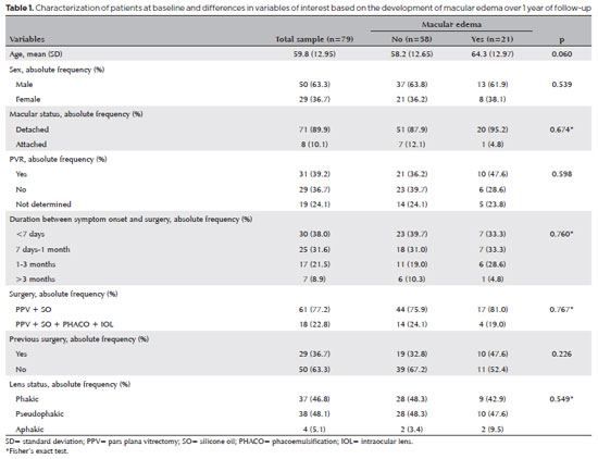
PURPOSE: To clarify the postoperative incidence of macular edema in patients undergoing surgery to repair rhegmatogenous retinal detachment and identify the associated risk factors.
METHODS: In this prospective, observational study, 79 patients who underwent surgery to correct rhegmatogenous retinal detachment using pars plana vitrectomy with silicone oil injection were analyzed. Patients were followed up postoperatively at 7, 30, 90, 180, and 365 days. At each visit, optical coherence tomography was performed to assess the presence or absence of macular edema. were analyzed as possible risk factors for macular edema: age, sex, macular status (attached or detached), presence of vitreoretinal proliferation, history of previous intraocular surgery, reported time of symptoms suggestive of rhegmatogenous retinal detachment up to the date of surgery, and the surgical modality performed.
RESULTS: The 1-year macular edema prevalence rate was 26.6%. In the adjusted analysis, older patients had a higher risk of macular edema, and each 1-year increase in age increased the risk of macular edema by 6% (95% confidence interval = 1.00-1.12). The macular status, vitreoretinal proliferation, the surgical technique used, prior intraocular surgery, and the intraocular lens status were not identified as risk factors. However, the incidence of macular edema increased up to 180 days after surgery, peaking at 10.6%, and then decreased until 365 days after surgery.
CONCLUSION: Macular edema was a common complication after surgery to treat rhegmatogenous retinal detachment, with its incidence peaking between 30 and 180 days after surgery. Age was an important risk factor for macular edema in this cohort.
Keywords: Macular edema; Retinal detachment; Vitrectomy; Tomography, optical coherence; Incidence; Risk factors
Arq. Bras. Oftalmol. 2025;88 (5 )
:1-8
| DOI: 10.5935/0004-2749.2024-0328
Abstract
PURPOSE: Posterior capsule rupture is defined as an intraoperative posterior capsule tear resulting in vitreous loss. This study aimed to analyze the clinical characteristics, preoperative risk factors, intraoperative management strategies, and postoperative complications associated with posterior capsule rupture during phacoemulsification surgery.
METHODS: This was a retrospective observational cohort study of the medical records for 25,224 phacoemulsification surgeries performed at our tertiary eye care center between 2017 and 2022. We collected and collated the demographic characteristics and clinical findings of the patients in our cohort. Intraoperative management strategies and postoperative outcomes over a 1-year followup period were also recorded.
RESULTS: Posterior capsule rupture occurred in 351 eyes (351 patients), giving an overall posterior capsule rupture rate of 1.3%. The mean patient age was 68.6 ± 10.8 years. Pseudoexfoliation syndrome, mature cataracts, brown cataracts, and surgery performed by a resident were identified as risk factors for posterior capsule rupture (p<0.05 for each; the risk ratios were 2.70, 2.15, 2.44, 1.34, respectively). The most common intraoperative complications were dislocated lens fragments in the vitreous (8%) and iris damage (7.1%). The mean best-corrected visual acuity improved from 1.31 ± 0.84 (logMAR) postoperatively to 0.51 ± 0.56 at the end of the 1-year follow-up period (p<0.001). Corneal edema (55.6%) and elevated intraocular pressure (33.3%) were the most common early postoperative complications. Persistently elevated intraocular pressure (11.1%) and cystoid macular edema (5.1%) were the most common late postoperative complications.
CONCLUSION: Posterior capsule rupture is a common complication of phacoemulsification surgery that requires prolonged postoperative follow-up and a multidisciplinary approach. Despite the increased incidence of complications when rupture occurs, appropriate intraoperative and postoperative management can lead to satisfactory visual outcomes.
Keywords: Cataract extraction; Phacoemulsification; Posterior capsule rupture; Corneal edema; Risk factors; Postoperative complications; Intraoperative complications


 ABO is licensed under a Creative Commons Attribution-NonComercial 4.0 Internacional.


08-tab01.jpg)
07-tab01.jpg)
08-tab01tb.jpg)
13-tab01tb.jpg)
13-fig01.jpg)

12-fig01.jpg)
12-fig01.jpg)
06-fig01.jpg)