Arq. Bras. Oftalmol. 2023;86 (4 )
:322-329
| DOI: 10.5935/0004-2749.20230058
Abstract
Introdução: Os oftalmologistas têm alto risco de contrair a doença do Coronavírus-19 devido à proximidade com os pacientes durante os exames com lâmpada de fenda. Usamos um modelo de computação para avaliar a eficácia das proteções para lâmpadas de fenda e propusemos uma nova proteção ergonomicamente projetada.
Métodos: As simulações foram realizadas no software comercial Star-CCM +. Os aerossóis de gotículas foram considerados 100% de água em fração de volume com distribuição de diâmetro de partícula representada por uma média geométrica de 74,4 ± 1,5 (desvio padrão) μm ao longo de uma duração de quatro minutos. A massa total de gotículas de água acumulada no manequim e a massa expelida pela boca do paciente foram medidas em três condições diferentes: 1) Sem protetor de lâmpada de fenda, 2) com protetor padrão, 3) Com o novo protetor proposto.
Resultados: A massa total acumulada das gotas de água (kg) e a porcentagem da massa expelida acumulada no escudo para cada uma das respectivas condições foram; 1) 5,84e-10 kg (28% do peso total da partícula emitida que assentou no manequim), 2) 9,14e-13 kg (0,045%), 3,19e-13 (0,015%). O escudo padrão foi capaz de proteger 99,83% das partículas que, de outra forma, teriam se depositado no manequim, o que é semelhante a 99,95% para o projeto proposto.
Conclusão: Protetores com lâmpada de fenda são ferramentas eficazes de controle de infecção contra gotículas respiratórias. O protetor proposto mostrou eficácia comparável em comparação com os protetores de lâmpada de fenda convencionais, mas potencialmente oferece uma melhor ergonomia para oftalmologistas durante o exame de lâmpada de fenda.
Keywords: Oftalmologistas; Infeções por coronavírus/prevenção & controle; Pandemias; Gotículas lipídicas; SARS-CoV-2; Lâmpada de fenda; Simulação por computador; Equipamentos de proteção; Desenho de equipamento.
Arq. Bras. Oftalmol. 2023;86 (3 )
:1-8
| DOI: 10.5935/0004-2749.20230034
Abstract
Purpose: To assess the outcomes of the trabecular bypass as replacement therapy for medications in pharmacologically controlled vs. pharmacologically uncontrolled open-angle glaucoma patients.
Methods: This was a retrospective study of eyes treated with first- (iStent) or second-generation (iStent inject) trabecular bypass. Group 1 consisted of eyes with pharmacologically controlled intraocular pressure <18 mmHg and Group 2 consisted of eyes with pharmacologically controlled intraocular pressure ≥18 mmHg. The main outcomes measured were qualified (with or without medications) and unqualified or complete (without medications) success rates at different target intraocular pressures, mean reduction (%) in medication use, and proportion of medication-free eyes.
Results: The mean age was 70.4 years in Group 1 (n=105) and 68.1 years in Group 2 (n=65). Qualified success rates for intraocular pressure <18 mmHg, intraocular pressure <15 mmHg, and intraocular pressure <12 mmHg were similar between the groups (Group 1: 96.2%, 88.6%, and 32.4%, respectively; Group 2: 93.8%, 78.5%, and 21.5%, respectively; all p>0.05). Complete success rates were significantly higher in Group 1 than in Group 2: for intraocular pressure <18 mmHg (76.2% vs. 47.7%), intraocular pressure <15 mmHg (73.3% vs. 40.0%), and intraocular pressure <12 mmHg (14.3% vs. 4.6%). The mean reduction in medication use was higher in Group 1 than in Group 2. At the end of follow-up, 79.0% of eyes in Group 1 and 47.7% of eyes in Group 2 became medication-free.
Conclusions: Both groups showed high qualified success rates, but eyes with baseline pharmacologically controlled intraocular pressure <18 mmHg showed higher complete success rates and greater chances of achieving no need for medications.
Keywords: Procedimentos cirúrgicos oftalmológicos; Extração de catarata; Glaucoma, ângulo aberto; Glaucoma/terapia; Glaucoma/cirurgia
Arq. Bras. Oftalmol. 2026;89 (1 )
:1-5
| DOI: 10.5935/0004-2749.2025-0045
Abstract
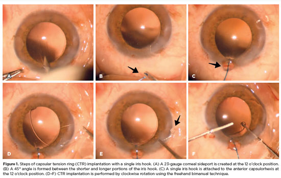
PURPOSE: To evaluate the effect of using a single iris retractor, affixed to the anterior capsulorhexis at the 12 o'clock position, on the ease of capsular tension ring implantation.
METHODS: This prospective comparative study comprised 37 patients with zonular weakness attributed to pseudoexfoliation syndrome who underwent capsular tension ring implantation during cataract surgery. In Group 1, a single iris retractor was inserted into the anterior capsulorhexis at the 12 o'clock position. Group 2 did not receive this intervention. Zonular weakness was graded on a scale of 1–5, and the subjective difficulty of capsular tension ring implantation was categorized as easy, medium, or difficult.
RESULTS: Group 1 and 2 comprised 20 and 17 patients, respectively. There were no significant differences between the groups in age, sex distribution, and presence of glaucoma (p=0.53, p=0.28, and p=1.00, respectively). The mean zonular weakness score was significantly higher in Group 1 (3.35 ± 0.45) than in Group 2 (2.71 ± 0.59; p=0.02). Capsular tension ring implantation was significantly easier in the iris retractor group (p<0.001).
CONCLUSIONS: Placement of a single iris retractor attached to the anterior capsulorhexis at the 12 o'clock position may facilitate easier capsular tension ring implantation, even in patients with greater zonular weakness. This technique could reduce the risk of capsular tension ring displacement into the iridocorneal angle or ciliary sulcus.
Keywords: Capsular tension ring; Cataract; Iris hook; Pseudoexfoliation syndrome; Zonular weakness; Cataract extraction; Phacoemulsification; Capsulorhexis.
Arq. Bras. Oftalmol. 2026;89 (2 )
:1-8
| DOI: 10.5935/0004-2749.2025-0175
Abstract
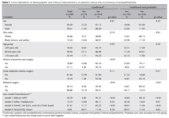
PURPOSE: Endophthalmitis is one of the most important adverse events after cataract surgery, as it can lead to total vision loss. This study aimed to describe the occurrence of endophthalmitis after phacoemulsification with intraocular lens implantation in patients treated in a community setting in Porto Velho, Rondônia, Brazil.
METHODS: This retrospective cohort study was conducted using a database of 649 medical records of patients who underwent surgery and were followed for three months. Poisson regression analysis was used to estimate relative risks and 95% confidence intervals (95% CIs).
RESULTS: The incidence of confirmed endophthalmitis was 11.94% (95% CI, 9.50-14.76), while the incidence of confirmed and probable cases was 20.50% (95% CI, 17.52-23.73). For confirmed cases, bilateral surgery and the use of lens model 3 were identified as risk factors for endophthalmitis, whereas age over 70 yr and preoperative antibiotic use were protective factors. For confirmed and probable cases, brown and yellow skin color, bilateral surgery, and the use of lens model 3 were also identified as risk factors. Gram-negative bacteria were the predominant etiological agents, and corneal edema was the main clinical manifestation. The mean duration of treatment was eight days, and 27.12% of patients used antibiotics.
CONCLUSION: The incidence observed was substantially higher than that reported in the literature, with a predominance of Gram-negative agents and an association with bilateral surgeries and the Eyeol intraocular lens model. These findings reinforce the need for continuous epidemiological surveillance and the implementation of specific biosafety and infection control protocols during cataract surgery campaigns.
Keywords: Endophthalmitis; Disease outbreaks; Phacoemulsification; Lens implantation, intraocular; Lenses, intraocular; Cataract; Risk factors; Anti-bacterial agents
Arq. Bras. Oftalmol. 2025;88 (4 )
:1-6
| DOI: 10.5935/0004-2749.2024-0249
Abstract
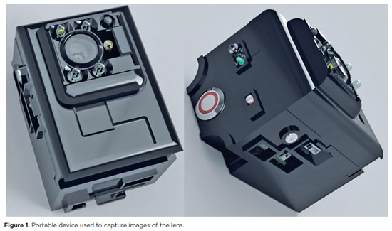
PURPOSE: Access to cataract treatment and diagnostic tools continues to be hindered by financial and logistical barriers. Thus, photography-based cataract analysis via portable devices offers a promising solution for the detection of cataracts in remote regions. In this study, the accuracy of a portable device that is based on the Lens Opacities Classification III System for diagnosing cataracts was analyzed.
METHODS: Photographs of the anterior segment of the eye were taken in a low-light environment, and the pupillary region markings were automatically delineated using infrared photography. The captured images were automatically analyzed using a convolutional neural network. The study group included patients with cataracts, and the control group included patients without cataracts.
RESULTS:A total of 270 eyes were analyzed, which included 143 eyes with cataracts and 127 control eyes. A total of 599 photos were analyzed. The isolated nuclear cataract was the most frequently detected subtype (37.5%), followed by a nuclear cataract associated with a cortical cataract (30.3%). The device's accuracy was 88.5% (Confidence intervals (CI), 83.19%–94.69%), specificity was 84.62% (CI 71.79%–97.30%), positive predictive value was 91.78% (CI 74.36%–97.30%), and negative predictive value was 82.50% (CI 74.36%–97.30%).
CONCLUSION: The portable device is a simplified user-friendly cataract screening technique that can interpret results in remote regions. This innovation could mitigate the occurrence of cataract-induced blindness and prevent premature surgical interventions in early-stage cataracts.
Keywords: Cataract/diagnosis; Diagnostic techniques ophthalmological/instrumentation; Optical devices; Equipment and supplies; Eye-tracking technology
Arq. Bras. Oftalmol. 2025;88 (6 )
:1-7
| DOI: 10.5935/0004-2749.2025-0083
Abstract
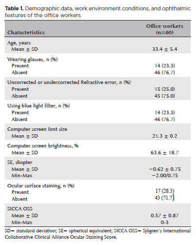
PURPOSE: To examine how ophthalmological features, screen exposure duration, and break habits among office employees affect ocular surface parameters.
METHODS: This single-center cross-sectional study involved two assessments on the same day: one before and one after a visual display terminal task. During the initial assessment, information on screen use was gathered, and refractive error, anterior segment examination, tear breakup time, and Schirmer test measurements were conducted. Participants tracked their screen usage and break durations throughout the day. At the end of the workday, tear breakup time and Schirmer I tests were repeated. Baseline and follow-up results were compared, and regression analysis was performed to identify factors linked to tear breakup time reduction.
RESULTS: The study enrolled 60 female office employees. Their mean screen time was 269.26 ± 70.21 min, with an average break duration of 151.93 ± 46.24 min. Tear breakup time at the second assessment (6.38 ± 2.70) was significantly lower than at baseline (8.62 ± 2.73) (p<0.001), whereas Schirmer test scores showed no significant change (p>0.05). Tear breakup time reduction was noted in 54 participants (90.0%), with a significant association between tear breakup time decrease percentage and screen exposure (p=0.001, r=0.463). Regression analysis showed that uncorrected or undercorrected refractive error was an independent risk factor for a ≥30% tear breakup time reduction, while taking more frequent short breaks (<15 min) acted as a protective factor.
CONCLUSIONS: Taking more frequent short breaks (<15 min) and correcting refractive errors help prevent intra-day tear breakup time decline during visual display terminal use. Structuring breaks to support tear film stability is advisable for occupations that require regular visual display terminal tasks.
Keywords: Tear film; Screen time; Tear breakup time; Office workers; Protective factors; Lacerations; Refractive errors; Risk factors.
Arq. Bras. Oftalmol. 2025;88 (6 )
:1-8
| DOI: 10.5935/0004-2749.2024-0394
Abstract
The advantages and disadvantages of using perioperative subconjunctival steroid injections in dropless cataract surgery continue to be debated. A systematic review of PubMed, EMBASE, and the Cochrane Central database identified five studies—two randomized controlled trials and three non-randomized studies—encompassing 70,751 eyes. Among these, 12,319 eyes (17.4%) received subconjunctival steroid injections, while 58,432 eyes (82.6%) were managed with topical steroids. The Cochrane Collaboration’s RoB 2 tool was applied for bias assessments in randomized controlled trials, and heterogeneity was assessed using the I² statistics. No statistically significant differences were found between the two groups regarding macular edema (p=0.249), visual acuity (p=0.73), or laser flare count (p=0.45). Both subconjunctival injections and topical steroids demonstrated comparable efficacy and safety in controlling postoperative inflammation after cataract surgery. Additional research is warranted to validate these conclusions.
Keywords: Cataract extraction; Phacoemulsification; Lens implantation, intraocular; Postoperative care; Intravitreal injections; Anti-inflammatory agents, non-steroidal/administration & dosage; Glucocorticoids; Triamcinolone acetonide; Research design; Randomiz
Arq. Bras. Oftalmol. 2025;88 (5 )
:1-8
| DOI: 10.5935/0004-2749.2024-0328
Abstract
PURPOSE: Posterior capsule rupture is defined as an intraoperative posterior capsule tear resulting in vitreous loss. This study aimed to analyze the clinical characteristics, preoperative risk factors, intraoperative management strategies, and postoperative complications associated with posterior capsule rupture during phacoemulsification surgery.
METHODS: This was a retrospective observational cohort study of the medical records for 25,224 phacoemulsification surgeries performed at our tertiary eye care center between 2017 and 2022. We collected and collated the demographic characteristics and clinical findings of the patients in our cohort. Intraoperative management strategies and postoperative outcomes over a 1-year followup period were also recorded.
RESULTS: Posterior capsule rupture occurred in 351 eyes (351 patients), giving an overall posterior capsule rupture rate of 1.3%. The mean patient age was 68.6 ± 10.8 years. Pseudoexfoliation syndrome, mature cataracts, brown cataracts, and surgery performed by a resident were identified as risk factors for posterior capsule rupture (p<0.05 for each; the risk ratios were 2.70, 2.15, 2.44, 1.34, respectively). The most common intraoperative complications were dislocated lens fragments in the vitreous (8%) and iris damage (7.1%). The mean best-corrected visual acuity improved from 1.31 ± 0.84 (logMAR) postoperatively to 0.51 ± 0.56 at the end of the 1-year follow-up period (p<0.001). Corneal edema (55.6%) and elevated intraocular pressure (33.3%) were the most common early postoperative complications. Persistently elevated intraocular pressure (11.1%) and cystoid macular edema (5.1%) were the most common late postoperative complications.
CONCLUSION: Posterior capsule rupture is a common complication of phacoemulsification surgery that requires prolonged postoperative follow-up and a multidisciplinary approach. Despite the increased incidence of complications when rupture occurs, appropriate intraoperative and postoperative management can lead to satisfactory visual outcomes.
Keywords: Cataract extraction; Phacoemulsification; Posterior capsule rupture; Corneal edema; Risk factors; Postoperative complications; Intraoperative complications
Arq. Bras. Oftalmol. 2025;88 (5 )
:1-7
| DOI: 10.5935/0004-2749.2024-0368
Abstract
PURPOSE: To compare endothelial corneal cell changes following cataract surgery performed by phacoemulsification with intraocular lens implantation, conducted by surgeons with varying levels of experience.
METHODS: Two hundred and eighty-three eyes diagnosed with cataract were included. Lens opacity was classified into three categories (I, II, and III). Surgeons were categorized into four experience levels (1, 2, 3, and 4), based on years of practice and lifetime surgeries performed. Corneal endothelial characteristics were assessed using non-contact specular microscopy, with measurements taken before surgery and 30-60 days post-surgery.
RESULTS: Pre- and postoperative endothelial analysis showed no significant differences between surgeon levels regarding visual acuity achieved, corneal thickness, and endothelial hexagonality. However, the central endothelial cell density index showed a significantly greater reduction among level 1 surgeons (p=0.026). Grade II cataracts exhibited significant variations in the central endothelial cell density (p=0.011) and average cell size, with level 1 surgeons showing the largest increases (p=0.024).
CONCLUSIONS: The analysis revealed significant differences in visual acuity and endothelial indices between surgeon experience levels, with less experienced surgeons showing greater variations and poorer performance. Clinical protocols should consider these data to establish safer training protocols.
Keywords: Cataract extraction; Phacoemulsification; Endothelium; corneal; Lens implantation, intraocular; Visual acuity; Internship and residency; Surgeons
Arq. Bras. Oftalmol. 2025;88 (3 )
:1-5
| DOI: 10.5935/0004-2749.2024-0084
Abstract
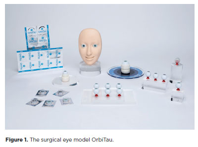
PURPOSE: The OrbiTau surgical simulator is a synthetic eye model developed to enhance cataract surgical training. Herein, we aimed to describe the perspectives of Harvard’s Ophthalmology faculty and residents regarding the effectiveness of OrbiTau.
METHODS: A cross-sectional study was conducted in which 11 surgeons from the Massachusetts Eye and Ear Infirmary, with prior experience utilizing simulated phacoemulsification platforms, conducted cataract surgery with the OrbiTau. Subsequently, they completed a satisfaction questionnaire using the Likert scale.
RESULTS: Regarding the various OrbiTau components, 90.90% of the participants reported that the OrbiTau lens capsule was comparable to that of the human lens during capsulotomy. Furthermore, 72.72% of the participants found that the OrbiTau lens consistency was analogous to that of the human lens nucleus. Approximately 63.63% of the participants reported that the model’s posterior lens capsule resembled the native posterior capsule, and 72.72% of the participants noted that the model’s red reflex was similar to that of the dilated human pupil. Most participants believed that the OrbiTau was easier to use and more realistic than other commercially available simulators.
CONCLUSION: Our single-institution survey of the Orbitau demonstrated that this model realistically replicates ocular structures and may be a viable option for cataract surgery training.
Keywords: Cataract extraction/education; Simulation training/methods; Ophthalmology/education; Phacoemulsification/education; Ophthalmologists/education; Surgeons/education; High fidelity simulation training
Arq. Bras. Oftalmol. 2024;87 (3 )
:1-5
| DOI: 10.5935/0004-2749.2023-0038
Abstract
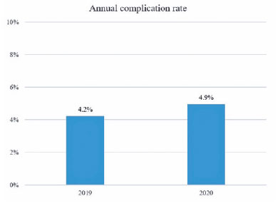
PURPOSE: To assess the effect of the coronavirus disease 2019 (COVID-19) pandemic on cataract surgery by residents who had mandatory surgical simulator training during residency.
METHODS: In this retrospective, observational analytical study, the total number of cataract surgeries and surgical complications by all senior residents of 2019 (2019 class; prepandemic) and 2020 (2020 class; affected by the reduced number of elective surgeries due to the COVID-19 pandemic) were collected and compared. All residents had routine mandatory cataract surgery training on a virtual surgical simulator during residency. The total score obtained by these residents on cataract challenges of the surgical simulator was also evaluated.
RESULTS: The 2020 and 2019 classes performed 1275 and 2561 cataract surgeries, respectively. This revealed a reduction of 50.2% in the total number of procedures performed by the 2020 class because of the pandemic. The incidence of surgical complications was not statistically different between the two groups (4.2% in the 2019 class and 4.9% in the 2020 class; p=0.314). Both groups also did not differ in their mean scores on the simulator’s cataract challenges (p<0.696).
CONCLUSION: Despite the reduction of 50.2% in the total number of cataract surgeries performed by senior residents of 2020 during the COVID-19 pandemic, the incidence of surgical complications did not increase. This suggests that surgical simulator training during residency mitigated the negative effects of the reduced surgical volume during the pandemic.
Keywords: COVID-19; Pandemics; Cataract extraction/education; Internship and residency/methods; Simulation training/methods; Phacoemulsification/education; Surgery, computer-assisted; Computer simulation; Clinical competence; Ophthalmology/education
 ABO is licensed under a Creative Commons Attribution-NonComercial 4.0 Internacional.
13-for01.jpg)
14-tab01.jpg)


15-fig01.jpg)
12-fig01.jpg)
06-fig01.jpg)