Arq. Bras. Oftalmol. 2023;86 (5 )
:1-6
| DOI: 10.5935/0004-2749.20230064
Abstract
Objetivo: Avaliar a resposta tecidual e clínica a um implante orbitário de polimetilmetacrilato, oco e multiperfurado em sua porção posterior em modelo animal após evisceração.
Métodos: Dezesseis coelhos da raça Nova Zelândia foram submetidos à evisceração do globo ocular direito. Todos receberam implante oco de polimetilmetacrilato de 12 mm de diâmetro, multiperfurado em sua semiesfera posterior. O estudo foi dividido em avaliação clínica e histopatológica. A avaliação clínica foi diária até 14 dias pós-evisceração e, a cada sete dias, até completar 180 dias. Os animais foram divididos em grupos de quatro animais e cada um foi submetido à exenteração com 07, 30, 90 e 180 dias e depois à eutanásia. A análise histopatológica teve por fim caracterizar o padrão inflamatório, a estrutura do colágeno e o grau de neovascularização. Para isso, além da tradicional coloração pela hematoxilina-eosina, utilizou-se o corante Picrosirius Red (PSR) e imuno-histoquímica com o marcador CD 34.
Resultados: Não houve sinais de infecção, afinamento conjuntival ou escleral, exposição ou extrusão do implante em nenhum animal durante o estudo. Já no sétimo dia, o tecido neoformado migrou para dentro do implante formando uma rede fibrovascular através dos canais posteriores. A resposta inflamatória diminuiu ao longo do tempo avaliado e não foram encontradas células gigantes multinucleadas.
Conclusão: O implante analisado permite a sua integração aos tecidos orbitários pelo crescimento fibrovascular em seu interior. Os autores acreditam que este modelo de implante orbital pode fazer parte de testes com humanos.
Keywords: Implantes orbitários; Polimetilmetacrilato; Evisceração ocular; Anoftalmia; Procedimentos cirúrgicos oftalmológicos; Coelhos.
Arq. Bras. Oftalmol. 2025;88 (6 )
:1-7
| DOI: 10.5935/0004-2749.2025-0006
Abstract
PURPOSE: This study aimed to evaluate the practices employed by oculoplastic surgeons in the assessment and management of anophthalmic sockets and external ocular prostheses.
METHODS: Oculoplastic surgeons from two countries, who specialized in the management of anophthalmic sockets, participated in a web-based survey. Data collected included demographics, types of surgery, implant use, external ocular prostheses management (including fabrication and cleaning), complications encountered, and follow-up times. The frequencies and distributions of the responses were statistically analyzed.
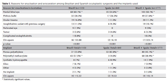
RESULTS: A total of 177 oculoplastic surgeons participated, 113 (63.8%) from Brazil, the remainder from Spain. Evisceration was the preferred surgical procedure of 149 (84.2%) surgeons. The most commonly reported indication for enucleation was a painful blind eye (n=103, 58.1%; both Brazil and Spain, p<0.001). Brazilian surgeons preferred polymethyl methacrylate implants (n=65, 57.5%), while Spanish surgeons favored porous polyethylene implants (n=53, 82.8%; p<0.001). Discharge was the most frequently observed clinical feature during socket evaluation (n=164, 92.6%; p<0.001). Brazilian surgeons recommended daily (n=53, 46.9%) or weekly (n=41, 36.2%) cleaning of external ocular prostheses, while Spanish surgeons more commonly recommended monthly cleaning (n=31, 48.4%; p<0.001). The majority of Brazilian surgeons (n=83, 73.4%) advised patients to remove their external ocular prostheses at night. Only a small number of Spanish surgeons (n=3, 4.6%) suggested this practice (p<0.001). Overall, the follow-up recommendations varied, with 70 (39.5%) surgeons recommending follow-up based on individual case needs, and 59 (33.3%) suggesting annual visits (p<0.001). The primary indications for external ocular prostheses replacement were edge damage (n=75, 42.3%) and loss of volume (n=68, 38.4%). The replacement intervals given typically ranged from 1 to 5 years (n=92, 51.9%; p<0.001).
CONCLUSION: Oculoplastic surgeons in Brazil and Spain demonstrated similar practices in the management of anophthalmic sockets. However, notable differences were observed in the choice of implant materials, cleaning protocols, and recommendations regarding external ocular prostheses removal during sleep.
Keywords: Anophthalmos; Eye, artificial; Polymethyl methacrylate; Polyethylene; Surgeons; Surveys and questionnaires; Brazil; Spain.
Arq. Bras. Oftalmol. 2026;89 (3 )
:1-6
| DOI: 10.5935/0004-2749.2025-0332
Abstract
PURPOSE: To quantitatively compare eyebrow and eyelid positions in anophthalmic sockets reconstructed with conical or spherical orbital implants combined with customized external ocular prostheses.
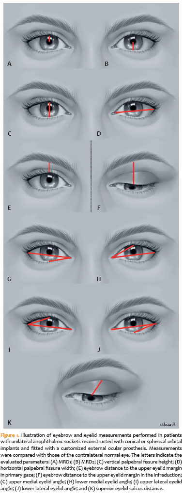
METHODS: This cross-sectional observational study included 38 patients with unilateral anophthalmic sockets, of whom 21 received conical implants, and 17 received spherical implants. Eyelid and eyebrow parameters—including margin reflex distance 1 and 2, vertical and horizontal palpebral fissure dimensions, eyebrow-to-upper-eyelid margin distance in primary gaze and infraduction, medial and lateral eyelid angles in primary gaze, and superior eyelid sulcus depth —were quantitatively assessed using standardized digital photographs analyzed with Image J software. The contralateral healthy eye served as the control. Statistical analyses were performed to compare measurements between groups.
RESULTS: In the primary gaze position, conical and spherical implants showed comparable margin-reflex distance1, margin-reflex distance2, vertical palpebral fissure height, eyelid margin position, and medial and lateral eyelid angles. During infraduction, the upper eyelid margin was significantly lower in sockets reconstructed with conical implants. Compared with contralateral normal eyes, anophthalmic sockets exhibited a reduced horizontal palpebral fissure and a deeper superior eyelid sulcus, irrespective of implant shape.
CONCLUSION: Anophthalmic sockets reconstructed with conical or spherical implants demonstrate similar eyebrow and eyelid positioning in primary gaze. However, conical implants are associated with a lower eyelid margin during infraduction. Independent of implant format, anophthalmic sockets show a narrower horizontal palpebral fissure and increased superior sulcus depth compared with normal eyes.
Keywords: Anophthalmos; Prosthesis implantation; Anophthalmic socket; Conical implants; Spherical implants; Orbital implants; Eyelid measurements
Arq. Bras. Oftalmol. 2024;87 (5 )
:1-7
| DOI: 10.5935/0004-2749.2023-0296
Abstract
PURPOSE: To compare inferomedial wall orbital decompression to balanced medial plus lateral wall orbital decompression in patients with Graves’ orbitopathy in the inactive phase with regard to exophthalmos reduction and the effects on quality of life.
METHODS: Forty-two patients with inactive Graves’ orbitopathy were randomly divided into two groups and submitted to one of two orbital decompression techniques: inferomedial wall orbital decompression or medial plus lateral wall orbital decompression. Preoperative and postoperative assessments included Hertel’s exophthalmometry and a validated Graves’ orbitopathy quality of life questionnaire. The results of the two groups were compared.
RESULTS: Compared to preoperative measurement, exophthalmos reduction was statistically significant in both groups (p<0.001) but more so in patients undergoing medial plus lateral wall orbital decompression (p=0.010). Neither orbital decompression techniques increased the visual functioning subscale score on the Graves’ orbitopathy quality of life questionnaire (inferomedial wall orbital decompression p=0.362 and medial plus lateral wall orbital decompression p=0.727), but a statistically significant difference was observed in the score of the appearance subscale in patients submitted to medial plus lateral wall orbital decompression (p=0.006).
CONCLUSIONS: Inferomedial wall orbital decompression is a good alternative for patients who do not require large exophthalmos reduction. However, medial plus lateral wall orbital decompression offers greater exophthalmos reduction and greater improvement in appearance (higher Graves’ orbitopathy quality of life questionnaire scores), making it a suitable option for esthetic-functional rehabilitation.
Keywords: Graves’ ophthalmopathy; Quality of life; Exophthalmos; Strabismus; Diplopia; Decompression, surgical
Arq. Bras. Oftalmol. 2024;87 (2 )
:1-8
| DOI: 10.5935/0004-2749.2022-0241
Abstract
PURPOSE: We aimed to study reported cases of nasopharyngeal carcinoma presenting with ophthalmic manifestations with and without a prior diagnosis of nasopharyngeal carcinoma.
METHODS: We conducted a systematic review following the Preferred Reporting Items for Systematic Reviews and Meta-Analyses (PRISMA). A literature search was conducted using the MEDLINE database in PubMed and Google Scholar. We included patients with a previous diagnosis of nasopharyngeal carcinoma in Group I and those without a prior diagnosis of nasopharyngeal carcinoma in Group II. Data included demographics, clinical presentation, history of nasopharyngeal carcinoma, treatment, histopathological description, World Health Organization classification, and outcome.
RESULTS: Fifty-eight patients (26 in Group I and 32 in Group II) were included. The male-to-female ratio was 3:1. The mean age of the patients (53.3 ± 11.7 years and 54.8 ± 16.2 years, respectively) and gender did not differ significantly between the two groups. The most common ocular presentations were diplopia and proptosis in the first group (each in 34.6%), whereas visual disturbance was most common in the second group (46.9%). Treatment options and World Health Organization grading were comparable. The outcome in 38 patients (after a comparable follow-up period) was significantly better in group II (p=0.003). There was no statistically significant difference in the outcome of 23 patients in correlation with World Health Organization grades II versus III irrespective of group (p=0.094).
CONCLUSIONS: The demographics of patients with nasopharyngeal carcinoma presenting with ophthalmic manifestations were similar between the two study groups, with a wide age range and male predominance. Patients presenting initially to ophthalmologists with no history of nasopharyngeal carcinoma have a more favorable outcome. World Health Organization grading may have less value as a prognostic indicator.
Keywords: Nasopharyngeal carcinoma; Carcinoma; Eye manifestations; Exophthalmos; Diplopia; Systematic review
 ABO is licensed under a Creative Commons Attribution-NonComercial 4.0 Internacional.
06-fig01.jpg)

02-fig01.jpg)
01-fig01.jpg)