Arq. Bras. Oftalmol. 2023;86 (4 )
:301-307
| DOI: 10.5935/0004-2749.20230054
Abstract
Objetivo: Avaliar os resultados visuais, satisfação e qualidade de vida de pacientes atendidos em um hospital escola pelo Sistema Único de Saúde, submetidos a implante bilateral de lente intraocular multifocal difrativa.
Métodos: Estudo tipo série de casos com intervenção, incluindo 20 pacientes submetidos a implante bilateral da lente intraocular multifocal difrativa EyeDiff® (Eyeol UK, Dunstable, UK). Os critérios de exclusão foram astigmatismo corneano >1,5 dioptria cilíndrica, cirurgia ou doença ocular prévias e complicações intraoperatórias ou pós-operatórias. Os pacientes foram avaliados após 1, 3 e 6 meses da cirurgia. Foram avaliadas a acuidade visual monocular e binocular para longe, intermediário e perto sob condições fotópica e mesópica, sensibilidade ao contraste monocular sob condições fotópicas, curva de defocus e questionário para avaliação da qualidade de vida.
Resultados: A acuidade visual para longe corrigida monocular foi de 0,3 logMAR ou melhor e a acuidade visual para perto com correção para longe foi J3 ou melhor em todos os olhos, sob condições fotópicas. A acuidade visual binocular para perto com a correção para longe foi J1 em todos os casos. A sensibilidade ao contraste estava no nível mínimo de normalidade para frequências espaciais baixas e altas e abaixo dos limites normais para frequência espacial intermediária. O questionário de qualidade de vida mostrou que os pacientes apresentavam altos níveis de satisfação.
Conclusão: O implante bilateral da lente intraocular multifocal EyeDiff® proporcionou boa acuidade visual e qualidade de vida, e independência de óculos aos pacientes. A acuidade visual e a sensibilidade ao contraste melhoraram progressivamente entre um e seis meses de pós-operatório.
Keywords: Acuidade visual; Qualidade de vida; Satisfação do paciente; Implante de lente intraocular; Sistema Único de Saúde.
Arq. Bras. Oftalmol. 2025;88 (6 )
:1-5
| DOI: 10.5935/0004-2749.2025-0085
Abstract
PURPOSE: The purpose of this study was to assess visual outcomes and patient satisfaction following cataract surgery involving the implantation of quad-loop intraocular lenses, including trifocal, bifocal, and toric variants.
METHODS: Information was obtained from both physical and electronic medical records of patients who underwent phacoemulsification cataract surgery with implantation of different intraocular lenses between January 1, 2022, and December 31, 2023. The study included individuals aged over 18 who received bilateral implantation of bifocal, trifocal, or monofocal toric intraocular lenses. Visual acuity was assessed at various postoperative time points using the logMAR scale. Quantitative variables were analyzed using mean and standard deviation.
RESULTS: A total of 92 eyes received premium intraocular lenses: 4 bifocal, 32 trifocal, 52 toric monofocal, and 4 trifocal toric lenses. The average preoperative corrected visual acuity was logMAR 0.478 ± 0.259. On the first postoperative day, the average uncorrected visual acuity was logMAR 0.301 ± 0.207. By day 30, 67.4% of eyes achieved uncorrected distance visual acuity of logMAR 0.2 or better. Patient satisfaction was high, with few reports of glare or halos.
CONCLUSION: Quad-loop intraocular lenses-including trifocal, bifocal, and toric models-demonstrated effective improvement in visual acuity and high levels of patient satisfaction. These lenses represent a suitable option for enhancing visual outcomes after cataract surgery. Additional studies with larger cohorts are recommended to confirm these results.
Keywords: Cataract extraction; Aberrometry/methods; Lenses, intraocular; Lens implantation, intraocular; Prosthesis design
Arq. Bras. Oftalmol. 2026;89 (2 )
:1-8
| DOI: 10.5935/0004-2749.2025-0175
Abstract
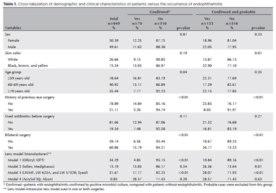
PURPOSE: Endophthalmitis is one of the most important adverse events after cataract surgery, as it can lead to total vision loss. This study aimed to describe the occurrence of endophthalmitis after phacoemulsification with intraocular lens implantation in patients treated in a community setting in Porto Velho, Rondônia, Brazil.
METHODS: This retrospective cohort study was conducted using a database of 649 medical records of patients who underwent surgery and were followed for three months. Poisson regression analysis was used to estimate relative risks and 95% confidence intervals (95% CIs).
RESULTS: The incidence of confirmed endophthalmitis was 11.94% (95% CI, 9.50-14.76), while the incidence of confirmed and probable cases was 20.50% (95% CI, 17.52-23.73). For confirmed cases, bilateral surgery and the use of lens model 3 were identified as risk factors for endophthalmitis, whereas age over 70 yr and preoperative antibiotic use were protective factors. For confirmed and probable cases, brown and yellow skin color, bilateral surgery, and the use of lens model 3 were also identified as risk factors. Gram-negative bacteria were the predominant etiological agents, and corneal edema was the main clinical manifestation. The mean duration of treatment was eight days, and 27.12% of patients used antibiotics.
CONCLUSION: The incidence observed was substantially higher than that reported in the literature, with a predominance of Gram-negative agents and an association with bilateral surgeries and the Eyeol intraocular lens model. These findings reinforce the need for continuous epidemiological surveillance and the implementation of specific biosafety and infection control protocols during cataract surgery campaigns.
Keywords: Endophthalmitis; Disease outbreaks; Phacoemulsification; Lens implantation, intraocular; Lenses, intraocular; Cataract; Risk factors; Anti-bacterial agents
Arq. Bras. Oftalmol. 2026;89 (1 )
:1-6
| DOI: 10.5935/0004-2749.2025-0140
Abstract
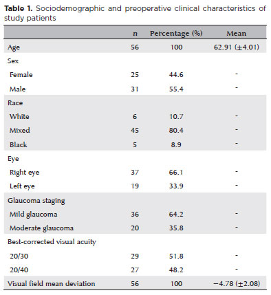
PURPOSE: Glaucoma is a chronic and progressive disease that requires long-term treatment and continuous monitoring. The Kahook Dual Blade, a device used to perform goniotomy in adults, is designed to improve intraocular pressure control in patients with glaucoma. This study aimed to evaluate the long-term efficacy and safety of kahook dual blade goniotomy in glaucoma patients undergoing cataract surgery over a 36-month follow-up.
METHODS: This was a retrospective case series including 56 eyes from 56 patients with mild-to-moderate primary open-angle glaucoma who underwent phacoemulsification combined with kahook dual blade goniotomy. Mean intraocular pressure values, number of preoperative and postoperative hypotensive eye drops, procedure survival, and complications were evaluated over 36 months. Surgical success was defined as either a reduction in intraocular pressure of ≥20% with intraocular pressure between 6 and 18 mmHg without additional medication or a reduction of ≥1 eye drop with intraocular pressure between 6 and
18 mmHg.
RESULTS: The mean preoperative intraocular pressure decreased from 15.96 ± 2,83) mmHg to 13.14 ± 2,11) mmHg at 36 months, representing a 14.9% reduction (p<0.001). The mean number of eye drops decreased from 1.91 ± 0,75) to 1.34 ± 0,92), a 29.8% reduction (p<0.001). The overall success rate was 69.6% at 36 months.
CONCLUSION: Kahook dual blade goniotomy combined with cataract surgery significantly reduced intraocular pressure and the number of hypotensive eye drops required in patients with mild-to-moderate primary open-angle glaucoma, with a favorable success rate maintained at 36 months.
Keywords: Glaucoma, open-angle/surgery; Gonioscopy/methods; Intraocular pressure/physiology; Lens implantation, intraocular; Phacoemulsification/methods; Trabeculectomy/instrumentation; Treatment outcome
Arq. Bras. Oftalmol. 2025;88 (4 )
:1-7
| DOI: 10.5935/0004-2749.2024-0190
Abstract
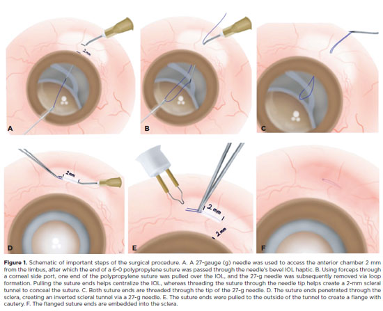
PURPOSE: The aim of this study is to describe a minimally invasive and atraumatic technique for managing the polypropylene suture-assisted scleral fixation of intraocular lens–capsular bag complex or artificial iris–intraocular lens complex for repositioning late luxated or subluxated intraocular lens–capsular bags and artificial iris–intraocular lens complexes.
METHODS: In this retrospective and observational study, we evaluated 11 patients, including 10 patients with capsular bag–intraocular lens complex subluxation or luxation into the vitreous cavity and 1 patient with an aniridia–intraocular lens complex. A single senior surgeon performed the procedures. After anesthesia, a 4 × 4 mm conjunctival peritomy was created, and a 6-0 polypropylene suture was passed through the sclera marked 2.0 mm posterior to the limbus. The suture ends were cauterized into a flange under 0.5 mm and inserted inversely into a scleral tunnel, concealed within a 2-mm scleral tunnel to ensure secure intraocular lens positioning.
RESULTS: We analyzed 11 patients with dislocated or dropped capsular bag–intraocular lens complexes. The patients' median age was 67 (range 44–78) years, with a median follow-up of 10 (range: 4–16) months. There were 8 (72%) men and 3 (27%) women. Conjunctival peritomy was performed in 4 (36%) patients. Predominantly, preoperative diagnoses indicated 7 (63%) patients with dislocated capsular bag–intraocular lens complexes. The capsular bag–intraocular lens complexes were centralized in all patients, and optical coherence tomography confirmed accurate suture positioning within the sclera. No suture-related complications were observed throughout the follow-up period, and no vision-threatening complications were reported during the postoperative follow-up.
CONCLUSIONS: Our technique provides a simple, effective solution for treating decentralized or dislocated capsular bag–intraocular lens complexes, eliminating the need for complex interventions such as large corneal wounds, scleral flaps, intraocular lens exchange, and intraocular lens externalization.
Keywords: Scleral fixation; Intraocular lens dislocation; Ophthalmologic surgical procedures; Sutures; Intraocular lens; Lens subluxation
Arq. Bras. Oftalmol. 2026;89 (3 )
:1-4
| DOI: 10.5935/0004-2749.2025-0263
Abstract
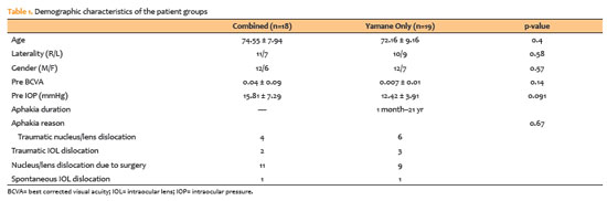
PURPOSE: To compare patients who underwent scleral fixation using the Yamane technique with and without simultaneous pars plana vitrectomy.
METHODS: A total of 37 patients were included in the study. Eighteen underwent simultaneous pars plana vitrectomy. The Yamane technique alone was performed only in patients with aphakia who had previously undergone pars plana vitrectomy for various reasons. Final lens position, best corrected visual acuity spherical equivalent, complication rates, and optical coherence tomography findings were recorded.
RESULTS: The duration of aphakia before intraocular lens implantation ranged from 1 month to 21 yr. Postoperative best corrected visual acuity improved in both groups, with no statistically significant difference (with pars plana vitrectomy: 0.42 ± 0.34; without pars plana vitrectomy: 0.32 ± 0.26; p=0.33). The spherical equivalent was also not significantly different between groups (with pars plana vitrectomy: 0.29 ± 1.08; without pars plana vitrectomy: 0.65 ± 2.23; p=0.53). There were no significant differences between the groups in complication rates, postoperative intraocular lens position or optical coherence tomography findings.
CONCLUSION: There was no difference in terms of safety or efficacy between the two approaches. Surgical decisions may be based on the surgeon’s experience and the patient’s systemic and ocular condition.
Keywords: Lens implantation, intraocular; Tomography, optical coherence; Vitrectomy; Intraocular lenses; Visual acuity; Aphakia; Yamane technique
Arq. Bras. Oftalmol. 2025;88 (5 )
:1-7
| DOI: 10.5935/0004-2749.2024-0368
Abstract
PURPOSE: To compare endothelial corneal cell changes following cataract surgery performed by phacoemulsification with intraocular lens implantation, conducted by surgeons with varying levels of experience.
METHODS: Two hundred and eighty-three eyes diagnosed with cataract were included. Lens opacity was classified into three categories (I, II, and III). Surgeons were categorized into four experience levels (1, 2, 3, and 4), based on years of practice and lifetime surgeries performed. Corneal endothelial characteristics were assessed using non-contact specular microscopy, with measurements taken before surgery and 30-60 days post-surgery.
RESULTS: Pre- and postoperative endothelial analysis showed no significant differences between surgeon levels regarding visual acuity achieved, corneal thickness, and endothelial hexagonality. However, the central endothelial cell density index showed a significantly greater reduction among level 1 surgeons (p=0.026). Grade II cataracts exhibited significant variations in the central endothelial cell density (p=0.011) and average cell size, with level 1 surgeons showing the largest increases (p=0.024).
CONCLUSIONS: The analysis revealed significant differences in visual acuity and endothelial indices between surgeon experience levels, with less experienced surgeons showing greater variations and poorer performance. Clinical protocols should consider these data to establish safer training protocols.
Keywords: Cataract extraction; Phacoemulsification; Endothelium; corneal; Lens implantation, intraocular; Visual acuity; Internship and residency; Surgeons
 ABO is licensed under a Creative Commons Attribution-NonComercial 4.0 Internacional.
10-fig01.jpg)

10-fig01.jpg)
13-fig01tb.jpg)
12-fig01.jpg)
03-fig01.jpg)
01-fig01.jpg)