Showing of 1 until 15 from 359 result(s)
Search for: Tomography, optical coherence; Ophthalmologic surgical procedures; Postoperative complications; Risk factors; Cataract; Cataract extraction; Low vision; Eye health
06-fig01.jpg)
Abstract
Objetivo: O objetivo deste estudo é comparar as medições de diâmetro corneano de dois dispositivos normalmente utilizados na prática clínica (IOL Master 500 e Atlas topógrafo corneal) para ver se são permutáveis. O fornecimento de informações sobre a permutabilidade de instrumentos poderia eliminar vários testes desnecessários e, consequentemente, reduzir a carga econômica para o paciente e para a sociedade.
Métodos: Nesta série de casos prospectivos e comparativos, a distância do diâmetro corneano foi medida por examinadores independentes utilizando o Topógrafo Atlas (Carl Zeiss Meditec) e o IOL Master 500 (Carl Zeiss Meditec), em um olho de 184 pacientes. A análise estatística foi realizada utilizando o teste t pareado, a correlação Pearson e a análise Bland-Altman para comparar os métodos de medição.
Resultados: As medições médias da distância do diâmetro corneano com o topógrafo Atlas e o IOL Master 500 foram de 12,20 ± 0,44 mm e 12,12 ± 0,41 mm, respectivamente (p<0,001). A diferença média de WTW entre os dois dispositivos foi de 0,07 mm (intervalo de confiança de 95% da diferença média: 0,04 - 0,11 mm). O coeficiente de correlação Pearson entre os dois dispositivos foi de 0,85, p<0,0001. Os limites de concordância de 95% entre os dois dispositivos foram de -0,38 mm a 0,53 mm.
Conclusões: O Atlas topographer e o IOL Master 500 podem ser utilizados permutavelmente em relação à medição do diâmetro corneano, uma vez que a gama de diferenças encontradas é pouco susceptível de afetar a prática clínica e a tomada de decisões.
Keywords: Topografia da córnea; Comprimento axial do olho; Técnicas de diagnóstico oftalmológico; Procedimentos cirúrgicos oftalmológicos.
15-tab01tb.jpg)
Abstract
Objetivo: Avaliar o implante de lente intraocular primária para tratamento da afacia pediátrica no Sistema Único de Saúde (SUS) e comparar os resultados em diferentes faixas etárias.
Métodos: Foram incluídas crianças com catarata congênita e do desenvolvimento unilateral ou bilateral de 0-12 anos de idade e submetidas a implante de lente intraocular primária.
Resultados: Cento e oito olhos de 68 crianças divididas em quatro grupos de idade (<7m; 7m-2a; 2-5a e > 5a) foram avaliados. Dezenove olhos (17,59%) apresentaram opacificação do eixo visual como complicação pós-operatória. Essa complicação foi mais frequente na faixa etária <7 meses (37,93%). A diferença foi significativa entre os grupos de idade <7 meses e > 5 anos (p=0,002). A opacificação do eixo visual foi dividida em duas categorias: membrana pupilar e proliferação de células do cristalino. Oito olhos apresentaram membrana pupilar e 14 proliferação de células do cristalino. Dos oito olhos com membrana pupilar, sete ocorreram na faixa etária <7 meses. A diferença entre o grupo de idade <7 meses e os grupos de 2-5 anos e > 5 anos foi significativa (p=0,01). A proliferação de células do cristalino foi mais frequente nos grupos de idade <7 meses e 2-5 anos, mas significativa apenas quando comparados o grupo de idade <7 meses com o grupo> 5 anos de idade (p=0,040). Glaucoma e suspeitos de glaucoma não foram observados durante o acompanhamento.
Conclusões: A principal complicação encontrada no estudo foi a opacificação do eixo visual. Sua incidência foi maior em crianças operadas antes dos 7 meses de idade.
Keywords: Extração de catarata; Lentes intraoculares; Complicações intraoperatórias; Glaucoma; Segmento anterior do olho; Criança.
14-tab01.jpg)
Abstract
Purpose: To assess the outcomes of the trabecular bypass as replacement therapy for medications in pharmacologically controlled vs. pharmacologically uncontrolled open-angle glaucoma patients.
Methods: This was a retrospective study of eyes treated with first- (iStent) or second-generation (iStent inject) trabecular bypass. Group 1 consisted of eyes with pharmacologically controlled intraocular pressure <18 mmHg and Group 2 consisted of eyes with pharmacologically controlled intraocular pressure ≥18 mmHg. The main outcomes measured were qualified (with or without medications) and unqualified or complete (without medications) success rates at different target intraocular pressures, mean reduction (%) in medication use, and proportion of medication-free eyes.
Results: The mean age was 70.4 years in Group 1 (n=105) and 68.1 years in Group 2 (n=65). Qualified success rates for intraocular pressure <18 mmHg, intraocular pressure <15 mmHg, and intraocular pressure <12 mmHg were similar between the groups (Group 1: 96.2%, 88.6%, and 32.4%, respectively; Group 2: 93.8%, 78.5%, and 21.5%, respectively; all p>0.05). Complete success rates were significantly higher in Group 1 than in Group 2: for intraocular pressure <18 mmHg (76.2% vs. 47.7%), intraocular pressure <15 mmHg (73.3% vs. 40.0%), and intraocular pressure <12 mmHg (14.3% vs. 4.6%). The mean reduction in medication use was higher in Group 1 than in Group 2. At the end of follow-up, 79.0% of eyes in Group 1 and 47.7% of eyes in Group 2 became medication-free.
Conclusions: Both groups showed high qualified success rates, but eyes with baseline pharmacologically controlled intraocular pressure <18 mmHg showed higher complete success rates and greater chances of achieving no need for medications.
Keywords: Procedimentos cirúrgicos oftalmológicos; Extração de catarata; Glaucoma, ângulo aberto; Glaucoma/terapia; Glaucoma/cirurgia
16-tab01tb.jpg)
Abstract
Objetivos: Rever características epidemiológicas de crianças submetidas a cirurgia de catarata, em centro de referência no estado de São Paulo, Brasil, e fatos associados a atrasos no tratamento.
Métodos: Um total de 240 olhos submetidos a cirurgia de catarata, em 178 crianças, foram revisados neste estudo transversal observacional. Os seguintes aspectos foram analisados: características clínicas e epidemiológicas, sinais apontados pelos pais, teste do reflexo vermelho, olho operado e idade no diagnóstico e na cirurgia.
Resultados: A média de idades na primeira visita e cirurgia de catarata foi de 48.9 meses (DP=50,0 meses) e 64.5 meses (DP=55.4 meses), respectivamente. O sinal mais importante apontado pelos pais foi a leucocoria. O teste do reflexo vermelho foi realizado em dois terços das crianças com resultados anormais em 28%. Histórico familiar de catarata foi evidente em 30 (20,9%) crianças (n=144). Os achados mais prevalentes em termos de histórico de problemas oculares foram: cirurgias oculares prévias em 37 (16,6%) olhos (n= 223), alterações do segmento anterior em 20 (9,0%) olhos (n=221), estrabismo em 21 (9,5%) olhos (n=220) e nistagmo em 38 (24,4%) crianças (n=156).
Conclusões: Uma das causas para o atraso na admissão pode ter sido a falha em realizar o teste do reflexo vermelho, apesar de não ter sido possível verificar se todas as crianças foram submetidas ao exame. A hereditariedade foi o fator mais importante quanto à causa da catarata nessas crianças. A presença de estrabismo e nistagmo mais uma vez aponta para o diagnóstico tardio. Ausência de programas de referência e centros oftalmológicos especializados em crianças, além do número restrito de profissionais de apoio treinados na área e especialistas em oftalmologia pediátrica, foram as barreiras mais importantes para o tratamento adequado da catarata em crianças.
Keywords: Catarata/ congênito; Extração de catarata; Técnicas de diagnóstico oftalmológico ; Baixa visão; Atenção terciária à saúde; Criança
Abstract
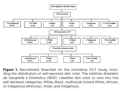
PURPOSE: This study evaluated macular thickness using spectral-domain optical coherence tomography in healthy participants from a population-based eye survey.
METHODS: The Brazilian Amazon Region Eye Survey was a population-based study assessing the prevalence and causes of visual impairment, blindness, and ocular diseases in adults aged ≥45 years from urban and rural areas of Parintins. A subgroup was selected based on inclusion criteria for both eyes: best-corrected visual acuity ≥20/32, normal eye examination results, and no prior ocular surgery. Scans were performed using the iVue optical coherence tomography device. Measurements were taken from the nine subfields defined by the Early Treatment Diabetic Retinopathy Study, examining the full retina as well as the inner and outer retinal layers. Associations of retinal thickness with age and sex were also analyzed. Statistical significance was set at p≤0.05.
RESULTS: In total, 70 healthy participants (25 males), aged 45–65 years (mean=52 ± 5), were included. Mean central foveal thickness was 248.71 ± 18.73 μm. A significant age-related reduction in macular thickness was observed, particularly in the inner superior parafovea (p=0.036), nasal perifovea (p=0.001), superior perifovea (p=0.028), outer layer of inferior parafovea (p=0.049), and the inferior perifovea of the full retina (p=0.029). Males showed significantly greater thickness in the outer layer, especially in the outer parafovea (p=0.004) and perifovea (p<0.0001).
CONCLUSIONS: This study established normative macular thickness values for healthy older adults in the Brazilian Amazon region using spectral-domain optical coherence tomography. Age and sex were found to significantly influence macular thickness and should be considered when interpreting measurements. These data will support future studies of retinal diseases in this population.
Keywords: Retinal diseases/diagnosis; Macula lutea/pathology; Macular degeneration/diagnosis; Diabetic retinopathy/diagnosis; Vision, low; Vision tests; Tomography, optical coherence/methods; Young adult; Cross-sectional studies; Brazil/epidemiology
Abstract
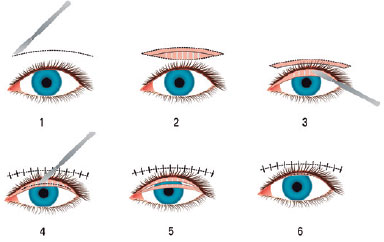
Objetivo: Compartilhar os resultados dos pacientes submetidos à rotação de retalho tarsal anterior, combinados com a reposição lamelar anterior devido à entrópio cicatricial da pálpebra superior e determinar a eficácia e a confiabilidade desta técnica cirúrgica.
Métodos: Foram incluídos neste estudo quinze olhos de 11 pacientes em quem realizamos cirurgia de rotação de retalho tarsal anterior combinada com reposição lamelar anterior devido ao entrópio cicatricial. Os registros médicos dos pacientes foram analisados retrospectivamente e as causas da entrópio cicatricial, bem como os achados do exame oftalmológico pré-operatório e pós-operatório foram registrados. A integridade anatômica e funcional da pálpebra foi considerada como sucesso cirúrgico.
Resultados: A idade média foi de 59,81 ± 18 anos. O período médio de seguimento foi de 21,72 ± 14 meses (intervalo 5-43 meses). As causas da entrópio cicatricial foram o desenvolvimento de cicatrizes pós-operatórias devido a eletrólises múltiplas para triquíase e/ou distiquiase em 7 olhos, tracoma em 6 olhos e trauma em 2 olhos. Todos os pacientes foram tiveram irritação e lacrimejamento pré-operatório, enquanto que 10 pacientes apresentavam opacidade e erosão da córnea e 1 paciente apresentava apenas erosão epitelial. O sucesso total anatômico e funcional foi alcançado em todos os casos.
Conclusão: A rotação do retalho tarsal anterior combinada com a reposição lamelar anterior no reparo da entrópio cicatricial é um procedimento cirúrgico alternativo efetivo e confiável.
Keywords: Tracoma/complicações; Pálpebras/cirurgia; Entrópio/cirurgia; Cicatriz; Retalhos cirúrgicos; Procedimentos cirúrgicos oftalmológicos/métodos
Abstract
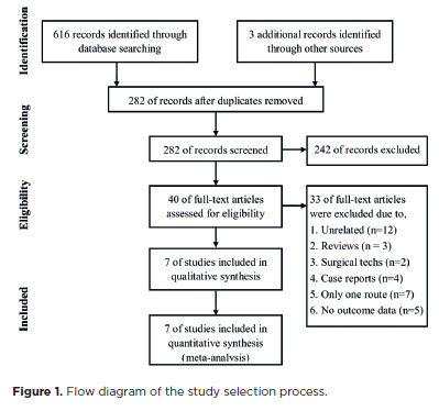
PURPOSE: To compare the incidence rates of complications following pediatric cataract surgery between the limbal and pars plana approaches.
METHODS: PubMed, EMBASE, Web of Science, Scopus, Cochrane Library, and ClinicalTrials.gov were systematically searched for studies comparing the two surgical approaches. We pooled the incidence rates of postoperative complications using a random-effects model.
RESULTS: Seven studies comprising 375 eyes from 260 patients were included. No significant differences in complication rates were observed between the limbal and pars plana approaches. The pooled incidence rates (95% confidence Interval) of postoperative visual axis opacity (VAO), VAO treated with laser or surgery, secondary glaucoma, wound leakage, corneal edema, anterior chamber reaction, posterior iris synechiae, capsular phimosis, intraocular lens dislocation, posterior capsular rupture, and intravitreal lens fragmentation were 4.7% (0.8%10.8%), 3.9% (1.0%-8.1%) , 2.8% (0%-11.4%), 0 (0%-1.3%), 2.9% (0%-11.8%), 5.6% (0.1%-16.5%), 2.4% (0%-8.5%), 3.8% (0.6%-8.9%), 2.2% (0%-6.4%), 9.2% (4.1%-15.8%) and 1.3% (0%-6.3%), respectively. Both surgical approaches demonstrated improved visual acuity postoperatively.
CONCLUSIONS: Pediatric cataract surgery, performed via the limbal or pars plana approach, is effective and safe, with a low incidence of complications when conducted by trained surgeons. Neither method demonstrated a significant difference in the visual acuity improvement or complication rates.
Keywords: Pediatric cataract surgery; Postoperative complications; Limbal route; Pars plana routes; Meta-analysis
Abstract
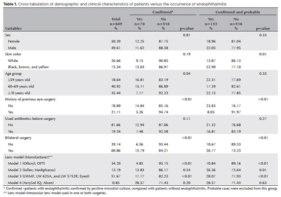
PURPOSE: Endophthalmitis is one of the most important adverse events after cataract surgery, as it can lead to total vision loss. This study aimed to describe the occurrence of endophthalmitis after phacoemulsification with intraocular lens implantation in patients treated in a community setting in Porto Velho, Rondônia, Brazil.
METHODS: This retrospective cohort study was conducted using a database of 649 medical records of patients who underwent surgery and were followed for three months. Poisson regression analysis was used to estimate relative risks and 95% confidence intervals (95% CIs).
RESULTS: The incidence of confirmed endophthalmitis was 11.94% (95% CI, 9.50-14.76), while the incidence of confirmed and probable cases was 20.50% (95% CI, 17.52-23.73). For confirmed cases, bilateral surgery and the use of lens model 3 were identified as risk factors for endophthalmitis, whereas age over 70 yr and preoperative antibiotic use were protective factors. For confirmed and probable cases, brown and yellow skin color, bilateral surgery, and the use of lens model 3 were also identified as risk factors. Gram-negative bacteria were the predominant etiological agents, and corneal edema was the main clinical manifestation. The mean duration of treatment was eight days, and 27.12% of patients used antibiotics.
CONCLUSION: The incidence observed was substantially higher than that reported in the literature, with a predominance of Gram-negative agents and an association with bilateral surgeries and the Eyeol intraocular lens model. These findings reinforce the need for continuous epidemiological surveillance and the implementation of specific biosafety and infection control protocols during cataract surgery campaigns.
Keywords: Endophthalmitis; Disease outbreaks; Phacoemulsification; Lens implantation, intraocular; Lenses, intraocular; Cataract; Risk factors; Anti-bacterial agents
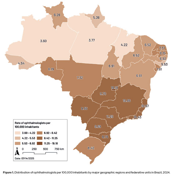
Abstract
PURPOSE: This study aimed to evaluate disparities in the distribution of ophthalmologists and the volume of cataract surgeries across Brazil, considering public and private health sectors and the country's federative units.
METHODS: Data on ophthalmologists were obtained from the National Medical Residency Commission and the Associação Múdica Brasileira. Information on cataract surgeries performed through the Unified Health System was collected from the DATASUS database, while data on procedures covered by private health plans were retrieved from the National Supplementary Health Agency. Population estimates from the 2024 Demographic Census of the Brazilian Institute of Geography and Statistics were used to calculate physician density and surgery rates per 100,000 inhabitants. Associations between the number of ophthalmologists and cataract surgery volume were analyzed using Spearman's correlation coefficient.
RESULTS: Brazil has 16,784 ophthalmologists, representing 8.96 specialists per 100,000 inhabitants. Marked disparities were observed: large cities (>500,000 inhabitants) had 18.75 ophthalmologists per 100,000 residents, whereas municipalities with <50,000 inhabitants had fewer than one. Across federative units, physician density ranged from 19.18 per 100,000 in the Federal District to 4.22 in Maranhão. In 2024, cataract surgery rates varied widely, from 1,012.61 per 100,000 inhabitants in the Southeast to 435.00 in the North. Nationally, Unified Health System performed 736.30 surgeries per 100,000 inhabitants, compared with 1,276.79 in the private sector. On average, each ophthalmologist performed 96.92 cataract surgeries annually.
CONCLUSION: Significant inequalities persist in the geographic distribution of ophthalmologists and in cataract surgery provision, with higher surgical volumes concentrated in the private sector. Targeted policies are required to address regional disparities and improve the equity and efficiency of cataract care delivery in Brazil.
Keywords: Ophthalmologists/supply & distribution; Ophthalmologists/statistics & numerical data; Cataract extraction; Health services accessibility/statistics & numerical data; Healthcare disparities; Health policy; Public health systems; Insurance, Heal
12-fig01tb.jpg)
Abstract
Objetivo: Comparar os achados oculares em longo prazo de crianças que se submeteram à cirurgia de catarata congênita antes dos dois anos de idade e receberam uma injeção intracameral de triancinolona no intraoperatório ou usaram prednisolona oral no pós-operatório para modular a inflamação ocular.
Métodos: Neste estudo prospectivo de coorte, todos os pacientes que participaram de um ensaio clínico anterior, que analisou os resultados cirúrgicos de 1 ano da cirurgia de catarata congênita usando triancinolona intracameral (Grupo de Estudo) ou prednisolona oral (Grupo Controle), eram elegíveis para participar. Os prontuários médicos dos pacientes foram revisados e as crianças foram submetidas a um exame oftalmológico completo no acompanhamento final. As principais medidas de desfecho foram: achados biomicroscópicos, pressão intraocular, espessura central da córnea, a necessidade de intervenções cirúrgicas adicionais e achados compatíveis com glaucoma.
Resultados: Vinte e seis olhos (26 pacientes) foram incluídos (Grupo de Estudo = 11 olhos; Grupo de Controle = 15 olhos). O seguimento médio foi de 8,2 ± 1,2 anos e 8,1 ± 1,7 anos nos Grupos de Estudo e Controle, respectivamente (p=0,82). Todos os olhos apresentavam lente intraocular centrada. Não houve diferença estatisticamente significativa entre os grupos com relação à presença de sinéquia posterior (p=0,56), pressão intraocular (p=0,49) ou espessura central da córnea (p=0,21). Nenhum dos olhos preencheu os critérios diagnósticos para glaucoma, apresentou opacificação secundária do eixo visual ou foi reoperado.
Conclusão: Os achados oculares em longo prazo de crianças que se submeteram à cirurgia de catarata congênita e receberam uma injeção intracameral de triancinolona no intraoperatório foram semelhantes aos que usaram prednisolona oral no pós-operatório para modular a inflamação ocular, sugerindo que a triancinolona intracameral pode substituir a prednisolona oral na cirurgia de catarata congênita, facilitando o tratamento pós-operatório e a adesão ao mesmo.
Keywords: Catarata congênita; Triancinolona; Prednisolona; Esteroides; Complicações pós-operatórias; Criança

Abstract
PURPOSE: This prospective, randomized, unmasked, clinical trial aimed to report the visual outcomes of cataract surgery on both eyes versus cataract surgery on one eye in Brazilian patients.
METHODS: This study included patients with bilateral cataracts and binocular visual acuity worse than or equal to 0.3 logarithm of the minimum angle of resolution. The patients were randomly assigned to undergo surgery on one (Control Group) or both eyes (one eye at a time; Intervention Group). Postoperatively, self-reported visual function using Catquest-9SF (primary outcome measure), binocular visual acuity, stereopsis, and ocular dominance (secondary outcome measures) were compared.
RESULTS: A total of 151 patients (77 and 148 eyes in the Control and Intervention Groups, respectively) completed the follow-up. Patients who underwent surgery on both eyes exhibited significantly better self-reported visual function (p=0.036) and stereopsis (p=0.026) than those who underwent surgery on one eye. Binocular visual acuity and ocular dominance did not affect the group comparisons.
CONCLUSIONS: Surgery on both eyes resulted in significantly better self-reported visual function and stereopsis than surgery on one eye.
Keywords: Cataract; Cataract extraction; Quality of life; Treatment outcome; Visual acuity; Binocular vision; Stereopsis
04-fig01tb.jpg)
Abstract
OBJETIVO: Descrever o processo de implementação e os resultados preliminares de um sistema de vigilância epidemiológica para endoftalmites associada à assistência à saúde.
MÉTODOS: Trata-se de um estudo de caso de implementação de um sistema de vigilância epidemiológica para endoftalmites. O sistema de vigilância epidemiológica para endoftalmites é um sistema estruturado que possibilita a vigilância de casos de endoftalmite associados à assistência à saúde após procedimentos oftalmológicos invasivos, desenvolvido e coordenado pela Divisão de Infecção Hospitalar da Secretaria de Estado da Saúde, São Paulo, Brasil. O processo de implementação incluiu uma fase piloto, seguida pela fase de expansão. Os dados foram enviados mensalmente à Divisão de Infecção Hospitalar pelos estabelecimentos de saúde participantes que realizaram procedimentos oftalmológicos no estado de São Paulo, Brasil no período de setembro de 2017 a dezembro de 2019.
RESULTADOS: Entre os 1.483 estabelecimentos de saúde elegíveis, 175 participaram do estudo (taxa de adesão de 11,8%), relatando 222.728 procedimentos oftalmológicos realizados, sendo 164.207 cirurgias de catarata e 58.521 injeções intravítreas. A taxa de incidência global de endoftalmite relatada foi de 0,05% (n=105; 80 casos após cirurgia de catarata e 25 casos após injeção intravítrea). As taxas de incidência entre os estabelecimentos de saúde variaram de 0,02% a 4,55%. A maioria dos casos foi causada por bactérias gram-positivas, principalmente Staphylococcus spp. Em 36 (46,2%) casos não houve crescimento bacteriano; nenhuma amostra foi coletada em 28 (26,7%) casos. O sistema de vigilância epidemiológica para endoftalmites possibilitou a identificação de um surto de quatro casos de endoftalmite após injeção intravítrea.
CONCLUSÃO: O sistema de vigilância epidemiológica para endoftalmites mostrou-se operacionalmente viável e eficiente para o monitoramento de casos de endoftalmite em nível estadual.
Keywords: Monitoramento epidemiológico; Endoftalmite; Atenção à saúde; Inquéritos epidemiológicos; Procedimentos cirúrgicos oftalmológicos

Abstract
PURPOSE: Posterior capsule rupture is defined as an intraoperative posterior capsule tear resulting in vitreous loss. This study aimed to analyze the clinical characteristics, preoperative risk factors, intraoperative management strategies, and postoperative complications associated with posterior capsule rupture during phacoemulsification surgery.
METHODS: This was a retrospective observational cohort study of the medical records for 25,224 phacoemulsification surgeries performed at our tertiary eye care center between 2017 and 2022. We collected and collated the demographic characteristics and clinical findings of the patients in our cohort. Intraoperative management strategies and postoperative outcomes over a 1-year followup period were also recorded.
RESULTS: Posterior capsule rupture occurred in 351 eyes (351 patients), giving an overall posterior capsule rupture rate of 1.3%. The mean patient age was 68.6 ± 10.8 years. Pseudoexfoliation syndrome, mature cataracts, brown cataracts, and surgery performed by a resident were identified as risk factors for posterior capsule rupture (p<0.05 for each; the risk ratios were 2.70, 2.15, 2.44, 1.34, respectively). The most common intraoperative complications were dislocated lens fragments in the vitreous (8%) and iris damage (7.1%). The mean best-corrected visual acuity improved from 1.31 ± 0.84 (logMAR) postoperatively to 0.51 ± 0.56 at the end of the 1-year follow-up period (p<0.001). Corneal edema (55.6%) and elevated intraocular pressure (33.3%) were the most common early postoperative complications. Persistently elevated intraocular pressure (11.1%) and cystoid macular edema (5.1%) were the most common late postoperative complications.
CONCLUSION: Posterior capsule rupture is a common complication of phacoemulsification surgery that requires prolonged postoperative follow-up and a multidisciplinary approach. Despite the increased incidence of complications when rupture occurs, appropriate intraoperative and postoperative management can lead to satisfactory visual outcomes.
Keywords: Cataract extraction; Phacoemulsification; Posterior capsule rupture; Corneal edema; Risk factors; Postoperative complications; Intraoperative complications
Abstract
PURPOSE: To clarify the postoperative incidence of macular edema in patients undergoing surgery to repair rhegmatogenous retinal detachment and identify the associated risk factors.
METHODS: In this prospective, observational study, 79 patients who underwent surgery to correct rhegmatogenous retinal detachment using pars plana vitrectomy with silicone oil injection were analyzed. Patients were followed up postoperatively at 7, 30, 90, 180, and 365 days. At each visit, optical coherence tomography was performed to assess the presence or absence of macular edema. were analyzed as possible risk factors for macular edema: age, sex, macular status (attached or detached), presence of vitreoretinal proliferation, history of previous intraocular surgery, reported time of symptoms suggestive of rhegmatogenous retinal detachment up to the date of surgery, and the surgical modality performed.
RESULTS: The 1-year macular edema prevalence rate was 26.6%. In the adjusted analysis, older patients had a higher risk of macular edema, and each 1-year increase in age increased the risk of macular edema by 6% (95% confidence interval = 1.00-1.12). The macular status, vitreoretinal proliferation, the surgical technique used, prior intraocular surgery, and the intraocular lens status were not identified as risk factors. However, the incidence of macular edema increased up to 180 days after surgery, peaking at 10.6%, and then decreased until 365 days after surgery.
CONCLUSION: Macular edema was a common complication after surgery to treat rhegmatogenous retinal detachment, with its incidence peaking between 30 and 180 days after surgery. Age was an important risk factor for macular edema in this cohort.
Keywords: Macular edema; Retinal detachment; Vitrectomy; Tomography, optical coherence; Incidence; Risk factors
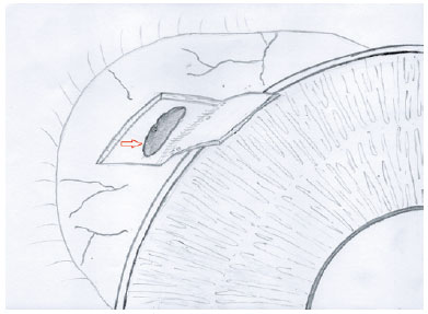
Abstract
The creation of a scleral flap during trabeculectomy can be complicated by a buttonhole, partial amputation at the limbus, and extensive thinning. In some cases, the procedure must be aborted to prevent more serious postoperative complications. This report describes a technique of converting complicated trabeculectomy into ab externo cyclodialysis. A 41-year-old patient with congenital glaucoma presented with a perforated scleral wall with the choroidal tissue exposed during the dissection of the partial-thickness scleral flap. By using a Barraquer cyclodialysis spatula through the scleral perforation, the choroid was separated from the sclera up to the scleral spur over 30º into the anterior chamber. The sclera and conjunctiva/Tenon were sutured with 10-0 nylon single sutures. Two months later, the intraocular pressure was reduced to 16 mmHg with no hypotensive topical medications. This case illustrates an alternative approach to managing a flap-related perioperative complication in trabeculectomy, which yielded good early results.
Keywords: Glaucoma/surgery; Trabeculectomy; Ophthalmologic surgical procedures /adverse effects; Cyclodialisys; Postoperative complications
 ABO is licensed under a Creative Commons Attribution-NonComercial 4.0 Internacional.
About
Issues
Editorial Board
Submission
Arquivos Brasileiros de Oftalmologia
Official publication of Brazilian Council of Ophthalmology - Conselho Brasileiro de Oftalmologia (CBO)
Rua Casa do Ator, 1.117 - 2nd floor - Zip Code: 04546-004
São Paulo - SP, Brazil
TEL: +55 11 3266-4000
E-mail: [email protected]