Arq. Bras. Oftalmol. 2023;86 (5 )
:1-5
| DOI: 10.5935/0004-2749.20230063
Abstract
Objetivo: A injeção peribulbar de triancinolona é um tratamento alternativo para doenças oculares da tireoide; no entanto, a segurança desse procedimento continua controversa. O objetivo deste artigo é descrever os efeitos adversos locais e sistêmicos de injeções peribulbares de triancinolona em pacientes com doença ocular da tireoide.
Métodos: Estudo retrospectivo de uma série de casos. Foram analisados os prontuários médicos dos pacientes com doença ocular da tireoide tratados com injeções de triancinolona peribulbar em uma única instituição acadêmica entre 2007 e 2019. Foram documentadas as complicações locais e sistêmicas.
Resultados: Um total de 123 pacientes foram tratados. Apenas 11 (8,9%) pacientes apresentaram complicações locais, sendo a mais frequente a presença de equimoses palpebrais superficiais (7,3%), enquanto 2 (1,6%) pacientes apresentaram complicações sistêmicas (hiperglicemia e inibição da suprarrenal após a interrupção do tratamento). Todas estas complicações foram transitórias e nenhum paciente apresentou sequelas de longo prazo.
Conclusões: As injeções peribulbares de triancinolona nas doenças oculares da tireoide têm uma taxa muito baixa de complicações, tanto locais quanto sistêmicas. São necessários estudos prospectivos para aprofundar este tópico.
Keywords: Órbita/diagnóstico por imagem; Imageamento por ressonância magnética; Oftalmopatia de Graves; Triancinolona/efeitos adversos; Injeções.
Arq. Bras. Oftalmol. 2023;86 (4 )
:359-364
| DOI: 10.5935/0004-2749.20230056
Abstract
Objetivo: Comparar as características radiológicas e clínicas do adenoma pleomórfico primário e do carcinoma adenoide cístico da glândula lacrimal.
Métodos: Este estudo revisou retrospectivamente os achados de imagem e os prontuários médicos de casos de adenoma pleomórfico e carcinoma adenoide cístico da glândula lacrimal.
Resultados: Foram avaliados 11 pacientes com adenoma pleomórfico e 16 pacientes com carcinoma adenoide cístico. Não houve diferenças estatisticamente significativas em relação à idade e sexo. Proptose foi o sintoma de apresentação mais comum em ambos os grupos. Os carcinomas adenoides císticos foram mais propensos que os adenomas pleomórficos a apresentarem massas palpáveis, diplopia, dor e perda sensorial, mas essa diferença entre os grupos não foi estatisticamente significativa. Não houve diferenças estatísticas em termos de homogeneidade e indentação do globo ocular entre os dois tipos de tumores em imagens de tomografia computadorizada (p>0,05). Também à tomografia computadorizada, a invasão óssea, a calcificação do tumor e o sinal em cunha foram mais frequentes nos carcinomas adenoides císticos, enquanto a remodelação óssea foi mais frequente nos adenomas pleomórficos, com significância estatística para todas essas manifestações (p<0,05). À ressonância magnética, os adenomas pleomórficos foram significativamente mais propensos a terem margens bem definidas, contornos lobulados, realce heterogêneo pelo contraste e hiperintensidade na ressonância magnética ponderada em T2 (p<0,05).
Conclusão: Ao se diferenciar o adenoma pleomórfico e o carcinoma adenoide cístico da glândula lacrimal, é muito importante avaliar as características radiológicas juntamente com as características clínicas. Os contornos lobulados podem ser uma característica radiológica significativamente distinta em favor do adenoma pleomórfico.
Keywords: Aparelho lacrimal/patologia; Adenoma pleomorfo; Carcinoma adenoide cístico; Tomografia computadorizada por raios x; Imagem por ressonância magnética.
Arq. Bras. Oftalmol. 2022;85 (2 )
:166-173
| DOI: 10.5935/0004-2749.20220034
Abstract
Objetivos: Mensurar a perfusão do complexo retina/coróide com ressonância magnética em olhos com fechamento angular primário agudo (FAPA).
Métodos: Três sequências de ressonância magnética, duas anatômicas e uma de perfusão com gadolínio, foram adquiridas em pacientes com fechamento angular primário agudo. Regiões de interesse foram desenhadas na sequência de perfusão e sobrepostas à sequência anatômica. O volume de sangue relativo nos 2 primeiros segundos foi considerado como referência, e sua variação nos 28 segundos subsequentes foi analisada.
Resultados: Cinco olhos de 5 pacientes com fechamento angular primário agudo foram incluídos (3 com crise unilateral e 2 com crise bilateral). Três olhos contralaterais e 2 olhos de 2 pacientes saudáveis, pareados por sexo e idade, foram incluídos no grupo controle. Pacientes com fechamento angular primário agudo incluíam 4 (80%) mulheres, com idade média de 65,8 ± 12,37 anos, pressão intraocular média de 56,2 ± 14,67 mmHg, pressão arterial média de 113,4 ± 8,17 mmHg e pressão de perfusão ocular de 57,2 ± 13,46mmHg. No grupo controle, pressão intraocular média foi de 15,6 ± 2,61 mmHg (p=0,0625), pressão arterial média de 107,4 ± 6,57 mmHg (p=1,00) e pressão de perfusão ocular de 91,8 ± 6,72 mmHg (p=0,0625). O volume de sangue relativo do complexo retina/coróide foi de -0,127 ± 0,048 nos olhos em fechamento angular primário agudo e -0,213 ± 0,116 nos olhos controles (p=0,3125).
Conclusões: A sequência de ressonância magnética com gadolínio não demonstrou diferença na perfusão de retina/coroide em olhos com fechamento angular primário agudo.
Keywords: Glaucoma de ângulo fechado; Imagem por ressonância magnética; Gadolínio; Retina; Perfusão
Arq. Bras. Oftalmol. 2025;88 (2 )
:1-7
| DOI: 10.5935/0004-2749.2023-0265
Abstract
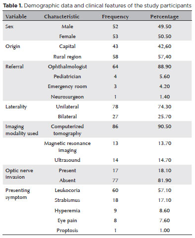
PURPOSE: Although Brazil has a high prevalence of retinoblastoma, there is a lack of epidemiological data on the disease. Thus, in this study, we aimed to evaluate the epidemiological profile of patients diagnosed with retinoblastoma in the ophthalmology department of a pediatric tertiary referral hospital in Ceara, Brazil.
METHODS: A descriptive and cross-sectional study was conducted by retrospectively analyzing the clinical and socioeconomic data from the medical records of pediatric patients followed-up at the hospital between 2007 and 2021. Retinoblastoma was diagnosed on the basis of a fundoscopic or histopathologic examination.
RESULTS: The data of 105 patients were included in the study, and the mean patient age at the time of diagnosis was 1.7 years. Most of the patients were women (50.5%) and hailed from rural areas (57.4%), which was associated with a higher tumor stage. Of the 150 patients, 57.1% initially presented with leukocoria. Ocular hyperemia was associated with more advanced stages of retinoblastoma (p=0.004). Bilateral involvement was observed in 25.7% of the patients and at a significantly younger age (p=0.009). The presence of retinal detachment, vascularized lesions, and vitreous seeds significantly increased the likelihood of requiring enucleation.
DISCUSSION: This study presents an epidemiological description of retinoblastoma in Brazil, which highlights the significance of early detection. Delayed diagnosis is associated with a poorer visual prognosis and higher mortality rate, particularly in patients with unilateral disease. Risk factors for a more severe disease were retinal detachment, vascularized lesions, and vitreous seeds. The correlation between histopathological features and clinical outcomes was limited.
CONCLUSION: Further studies are required to assess the influence of ocular hyperemia, fundoscopic assessment, and histopathologic findings on the prognosis of retinoblastoma. Moreover, it is critical to devise interventions to reduce the time-to-diagnosis in rural areas.
Keywords: Retinoblastoma; Retinal neoplasms; Epidemiology; Prevalence; Risk factors; Delayed diagnosis; Child
Arq. Bras. Oftalmol. 2025;88 (3 )
:1-8
| DOI: 10.5935/0004-2749.2024-0104
Abstract
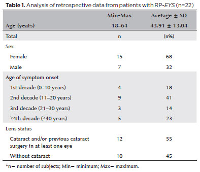
PURPOSE: This study aimed to characterize retinitis pigmentosa associated with the eyes shut homolog gene, which causes hereditary retinal degeneration.
METHODS: The anatomical and functional findings of retinitis pigmentosa in patients with variants of the eyes shut homolog gene were characterized and compared using multimodal imaging and genetic analysis of the variants. Clinical data such as visual acuity, lens status, and refraction were obtained from medical records. Patients underwent an ophthalmic examination, including static visual field, microperimetry, optical coherence tomography, fundus autofluorescence, and fundus photography.
RESULTS: Twenty-two patients were included in the study. Several anatomical and functional characteristics of retinitis pigmentosa-eyes shut homolog were identified, including the presence of cataracts, cystoid macular edema, epiretinal membrane, and a tubular visual field. Genetic results revealed 26 distinct variants in the cohort, with 7 novel variants not previously documented or reported in the scientific literature or databases.
CONCLUSION: The findings demonstrate that eyes shut homolog-retinitis pigmentosa manifests in specific patterns, starting in adolescence with mild progression and advancing with age. The integration of multimodal imaging and genetic analysis has provided a detailed understanding of the anatomical and functional features of retinitis pigmentosa-eyes shut homolog. Seven novel variants of the eyes shut homolog gene have been identified. These findings enhance the understanding of eyes shut homolog-related retinitis pigmentosa characteristics of by detailing the spectrum of mutations in this gene within the Brazilian population.
Keywords: Retinal diseases/diagnostic imaging; Retinitis pigmentosa/genetics; Retinal degeneration; Eye proteins/genetics; Eye diseases, hereditary/genetics; Genes, recessive; Phenotype; Multimodal imaging; Tomography, optical coherence/methods; Fluorescein angiogr
Arq. Bras. Oftalmol. 2026;89 (4 )
:1-5
| DOI: 10.5935/0004-2749.2025-0356
Abstract
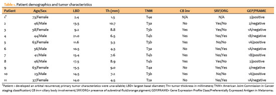
PURPOSE: To analyze the results of the 15-gene expression profile/preferentially expressed antigen in melanoma (15-GEPP) prognostic classifier and associated survival outcomes in a series of Brazilian patients with posterior uveal melanoma.
METHODS: Patients born and raised in Brazil who were diagnosed with posterior uveal melanoma and underwent fine-needle aspiration biopsy for 15-GEPP prognostic testing using the DecisionDx-Uveal melanoma and DecisionDx-PRAME assays between September 2020 and 2023 were included. All patients were diagnosed and treated by the senior author.
RESULTS: Of 236 patients diagnosed with posterior uveal melanoma during the study period, 11 (4.6%) met the inclusion criteria, including six males and five females. Based on 15-GEPP results, seven patients (63.6%) were classified as Class 1 and 4 (36.4%) as Class 2. Preferentially expressed antigen in melanoma (PRAME) expression was negative in six patients (54.6%) and positive in five patients (45.4%). During a mean follow-up of 36.4 ± 17.6 months, five patients developed metastasis: two with Class 2/PRAME-positive tumors, one with Class 2/PRAME-negative tumors, one with Class 1/PRAME-positive tumors, and one with Class 1/PRAME-negative tumors. Three of these patients died. The mean survival after the diagnosis of metastasis was 21.2 months (median: 17.0 months).
CONCLUSIONS: The proportions of Class 1 versus Class 2 tumors and PRAME-negative versus PRAME-positive tumors as well as the prognostic performance of these biomarkers appear similar in Brazilian patients with posterior uveal melanoma compared with large published cohorts from North America. Further studies with larger Brazilian cohorts are needed to validate these findings.
Keywords: Uveal melanoma; Fine-needle biopsy; Prognosis; Biomarkers; Neoplasms; Transcriptome; Survival; Brazil


 ABO is licensed under a Creative Commons Attribution-NonComercial 4.0 Internacional.


05-tab01.jpg)
11-tab01.jpg)
05-fig01.jpg)
10-fig01.jpg)
11-fig01tb.jpg)

03-fig01.jpg)
01-fig01.jpg)
14-fig01.jpg)
07-tab01tb.jpg)
05-fig01.jpg)