Arq. Bras. Oftalmol. 2022;85 (6 )
:584-589
| DOI: 10.5935/0004-2749.20220089
Abstract
Objetivo: Determinar os efeitos da deficiência de vitamina D nos microvasos da retina usando angiotomografia de coerência óptica.
Métodos: Este estudo foi planejado para ser do tipo caso-controle observacional. Foram avaliados 98 olhos de pacientes com deficiência de vitamina D e 96 olhos de participantes saudáveis com nível sérico de vitamina D superior a 30 ng/mL. Foram adquiridas imagens de varredura centralizadas na mácula, com um tamanho de 6,00 × 6,00 mm. Mediram-se a densidade dos vasos nos plexos capilares superficial e profundo da retina, a área da zona avascular foveal e a área do fluxo coriocapilar.
Resultados: Os grupos mostraram-se semelhantes em relação à melhor acuidade visual corrigida, ao gênero, ao comprimento axial, ao erro refrativo, à idade e à pressão intraocular ajustada. O nível médio de vitamina D foi significativamente menor no grupo de estudo (p=0,021). As densidades total, parafoveal e perifoveal do plexo capilar profundo foram significativamente maiores no grupo de estudo que no grupo controle (respectivamente, p=0,012, p=0,014 e p=0,023). As áreas da zona avascular foveal e do fluxo coriocapilar foram semelhantes nos dois grupos (respectivamente, p=0,37 e p=0,27). Além disso, houve uma forte correlação negativa do nível sérico de vitamina D com as densidades vasculares medidas em toda a imagem e nas regiões parafoveais e perifoveais do plexo capilar profundo no grupo de estudo (respectivamente, ρ de Spearman = -0,71, p=0,043; ρ de Spearman = -0,79, p=0,011; e ρ de Spearman = -0,74, p=0,032).
Conclusão: Pode ocorrer um aumento na densidade vascular da retina devido a alterações estruturais dos vasos causadas pela deficiência de vitamina D. O aumento da densidade vascular, especialmente no plexo capilar profundo, pode ser usado para o diagnóstico precoce da vasculopatia associada à deficiência de vitamina D.
Keywords: Deficiência de vitamina D; Vasos retinianos/fisiopatologia; Doenças vasculares/ prevenção & controle; Tomografia de coerência óptica
Arq. Bras. Oftalmol. 2022;85 (4 )
:370-376
| DOI: 10.5935/0004-2749.20220032
Abstract
Objetivos: Investigar a incidência, fatores de risco e desfechos visuais do desenvolvimento da membrana epirretiniana após reparo do descolamento regmatogênico da retina.
Métodos: Trata-se de um estudo retrospectivo de 309 olhos submetidos à cirurgia inicial para descolamento regmatogênico da retina primário sem complicações. Os exames foram realizados no pré-operatório aos 1, 3, 6 e 12 meses pós-operatórios. Os pacientes foram divididos em dois grupos, dependendo da presença ou ausência de membrana epirretiniana.
Resultados: A incidência de membrana epirretiniana pós-operatória foi de 28,5%; 42,7% desses pacientes apresentaram desenvolvimento grave da membrana epirretiniana e, portanto, foram submetidos à remoção desta membrana. A regressão logística mostrou que as lágrimas retinianas gigantes (RC: 2,66; 95% IC: 1,045 - 6,792, p=0,040) e lágrimas em ferradura (RC: 0,534; 95% IC: 0,295-0,967, p=0,039), foram preditores significativos de membrana epirretiniana pós-operatória. A coloração com acetonida de triancinolona foi significativamente associada à prevenção da membrana epirretiniana (p=0,022). Trinta e quatro pacientes apresentaram acuidade visual melhorada, ou igual, ou acuidade visual final melhor corrigida; 4 olhos foram avaliados na consulta final de acompanhamento e apresentaram redução da acuidade visual melhor corrigida.
Conclusão: Nossa análise demonstra que as lágrimas de ferradura e as lágrimas retinianas gigantes representam fatores de risco para a membrana epirretiniana pós-operatória. A coloração com acetonida de triancinolona teve um efeito preventivo significativo na membrana epirretiniana no pós-operatório. Além disso, uma segunda rodada de vitrectomia pars plana, incluindo remoção da membrana, levou a uma melhora significativa da acuidade visual final melhor corrigida na última consulta de acompanhamento, embora a recuperação tenha sido limitada.
Keywords: Membrana epirretiniana; Descolamento da retina; Recurvamento da esclera; Acuidade visual; Vitrectomia
Arq. Bras. Oftalmol. 2025;88 (3 )
:1-11
| DOI: 10.5935/0004-2749.2024-0049
Abstract
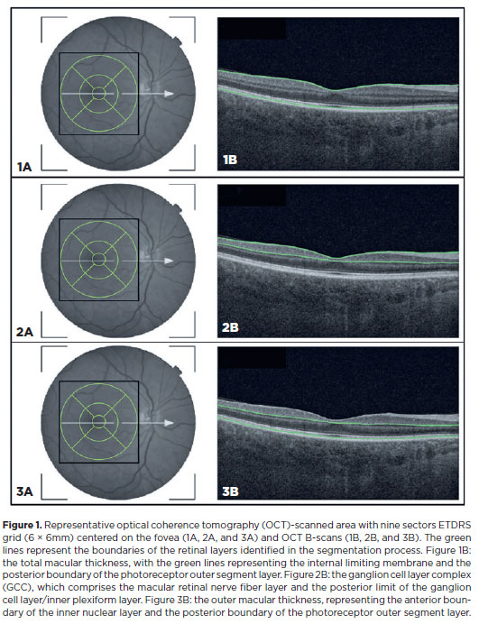
PURPOSE: This study aimed to evaluate the total macular thickness as well as the thickness of the inner and outer retinal layers in patients with Parkinson's disease. It also aimed to verify the correlation of these parameters with motor symptoms and cognitive function.
METHODS: A total of 46 eyes of 23 patients with Parkinson's disease and 40 eyes of 20 healthy controls were included in the study. The patients' cognitive, functional, and nonmotor symptoms were evaluated using the Katz Index of Independence and Pfeffer's Activities of Daily Living, Mini-Mental State Examination, Frontal Assessment Battery, Schwab and England Staging Scales, and Movement Disorders Society Nonmotor Symptoms Scale. The macular thickness measurements obtained via total, inner, and outer optical coherence tomography were recorded. Furthermore, the correlation of the parameters of optical coherence tomography with cognitive, functional, and nonmotor symptoms was assessed.
RESULTS: The scores of the Katz Index of Independence and Pfeffer's Activities of Daily Living as well as the Movement Disorders Society Nonmotor Symptoms Scale were significantly lower in patients with Parkinson's disease than in healthy controls. Moreover, the former had greater total macular thickness. The temporal and inferior outer sectors were significantly greater for the ganglion cell complex thickness in patients. A significant correlation was observed between the total macular thickness and the Movement Disorder Society-Unified Parkinson's Disease Rating Scale, Parte III (MDS-UPDRS-III) values. Contrarily, there was a negative correlation between the outer macular thickness and the MDS-UPDRS-III values. Meanwhile, the total macular thickness and ganglion cell complex thickness were significantly correlated with the scores of the Mini-Mental State Examination, Schwab and England Staging Scale, Frontal Assessment Battery, and Katz Index of Independence and Pfeffer's Activities of Daily Living. In addition, the Schwab and England scale was correlated with the outer macular thickness.
CONCLUSION: The total and inner macular thicknesses at the temporal and inferior outer sectors were greater in patients with Parkinson's disease than in the control group. These findings indicate that macular thickness may be greater in those with Parkinson's disease, particularly when associated with mild motor symptoms. In addition, the parameters of the total, inner, and outer optical coherence tomography were significantly associated with motor and nonmotor symptoms as well as cognitive function impairment.
Keywords: Parkinson's disease; Tomography, optical coherence; Neurodegenerative diseases; Cognitive dysfunction; Cognition; Motor perception; Visual acuity; Retina
Arq. Bras. Oftalmol. 2026;89 (4 )
:1-7
| DOI: 10.5935/0004-2749.2025-0344
Abstract
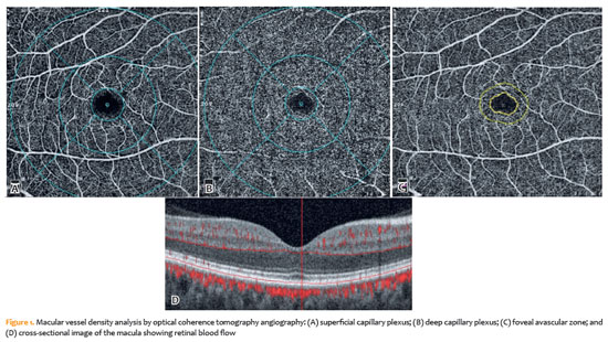
PURPOSE: This study aimed to evaluate early postoperative changes in retinal microvasculature in patients undergoing cardiopulmonary bypass surgery by comparing preoperative and postoperative measurements of superficial capillary plexus and, deep capillary plexus vessel density, foveal avascular zone area using optical coherence tomography angiography (OCT-A).
METHODS: This prospective longitudinal study included 38 patients, undergoing cardiopulmonary bypass surgery. Each patient served as their own control optical coherence tomography angiography imaging was performed at four time points: 3 days preoperatively and at postoperative week 1, week 2, and month 1. Vessel densities in the superficial capillary plexus and deep capillary plexuses and the foveal avascular zone area were measured and analyzed. Depending on data distribution repeated measures analysis of variance or the Friedman test was used for statistical analysis.
RESULTS: Data from all 38 patients were included in the analysis. A significant increase in overall superficial capillary plexus vessel density was observed following cardiopulmonary bypass surgery (p=0.008). In contrast, deep capillary plexus vessel density did not differ significantly between the preoperative and postoperative assessments. The foveal avascular zone area showed a significant increase at postoperative week 2 compared with baseline (p=0.016).
CONCLUSION: Cardiopulmonary bypass surgery may induce early retinal microvascular alterations, particularly in the superficial capillary plexus and foveal avascular zone. Optical coherence tomography angiography is a noninvasive and reproducible imaging modality that enables precise assessment of subtle retinal vascular changes after major cardiovascular surgery. These findings may contribute to postoperative monitoring and risk assessment in patients undergoing cardiopulmonary bypass.
Keywords: Cardiopulmonary bypass surgery; Extracorporeal circulation; Macular vessel density of the retina; Retina/blood supply; Foveal avascular zone; Optical coherence tomography angiography; Retinal microvasculature


 ABO is licensed under a Creative Commons Attribution-NonComercial 4.0 Internacional.


14-fig01.jpg)
03-tab01tb.jpg)
08-fig01.jpg)
13-fig01.jpg)
04-fig01.jpg)
03-fig01.jpg)
03-fig01.jpg)
15-fig01.jpg)
05-fig01.jpg)
02-fig01.jpg)