Arq. Bras. Oftalmol. 2023;86 (2 )
:113-120
| DOI: 10.5935/0004-2749.20230022
Abstract
Objetivos: Avaliar a estabilidade e eficácia da técnica double-flanged com sutura de 5-0 polipropileno para fixação de cataratas subluxadas aos 18 meses e as possíveis complicações desta nova técnica.
Métodos: Esta técnica utiliza um monofilamento de polipropileno 5-0 para criar dois flanges com um termocautério para fixar um Segmento de Tensão Capsular na esclera a fim de estabilizar o saco capsular subluxado. Esta técnica foi implementada em 17 olhos que necessitavam do implante de lente intraocular em casos de diálise zonular devido a trauma, síndrome de Marfan, microesferofacia, subluxação idiopática ou pós-facoemulsificação que provocou subulxação do saco capsular intraoperatória.
Resultados: O seguimento dos pacientes foi de 18 meses. A acuidade visual corrigida melhorou significativamente de 0,85 para 0,39 (logMAR), enquanto os erros de refração esféricos e cilíndricos e a pressão intraocular permaneceram estáveis. Nenhuma fotodegradação de sutura ou pseudofacodonese foi encontrada.
Conclusão: A técnica double-flanged para fixação transescleral de saco capsular com sutura de 5-0 polipropileno mostrou resultados de estabilidade de longo prazo para o complexo lente/saco capsular. Então, aparenta ser uma opção segura para cirurgia de catarata, sem necessidade pontos, em olhos com fraqueza zonular ou diálise
Keywords: Catarata; Facoemulsificação; Lente intraocular; Técnica de sutura; Acuidade visual
Arq. Bras. Oftalmol. 2021;84 (6 )
:561-568
| DOI: 10.5935/0004-2749.20210093
Abstract
Objetivo: Fornecer orientações sobre a frequência e os componentes dos exames oftalmológicos para crianças saudáveis de 0 a 5 anos.
Métodos: Essas diretrizes foram desenvolvidas com base em revisão bibliográfica e experiência clínica de um comitê de especialistas. Foram realizadas buscas PubMed/Medline; documentos selecionados não se restringiram a revisões sistemáticas, ensaios clínicos randomizados e estudos observacionais. Quando adequado, o perfil GRADE foi aplicado para graduá-los e o consenso de especialistas foi usado nos tópicos sem evidência científica. Também foram revisadas as recomendações pela Academia Americana de Pediatria, Associação Americana de Oftalmologia Pediátrica e Estrabismo, Academia Americana de Oftalmologia, Royal College of Ophthalmologist e Sociedade Canadense de Oftalmologia. O documento final foi aprovado pela Sociedade Brasileira de Oftalmologia Pediátrica e Sociedade Brasileira de Pediatria.
Resultados: Os recém-nascidos devem ser submetidos ao teste do reflexo vermelho e inspeção dos olhos e anexos pelo pediatra dentro de 72 horas de vida ou antes da alta da maternidade. O teste do reflexo vermelho deve ser repetido pelo pediatra durante as consultas de puericultura pelo menos três vezes ao ano durante os primeiros 3 anos de vida. Se factível, um exame oftalmológico completo pode ser feito entre 6 a 12 meses de vida. Até os 36 meses de idade, os marcos visuais, função visual apropriada para a idade, fixação e alinhamento ocular também devem ser avaliados pelo pediatra ou médico da família. Pelo menos um exame oftalmológico completo deve ser realizados entre 3 e 5 anos de idade. O exame deve conter pelo menos inspeção dos olhos e anexos, avaliação da função visual apropriada para a idade, avaliação da motilidade e alinhamento ocular (testes de cobertura), refração sob cicloplegia e avaliação do fundo de olho dilatado.
Conclusões: As diretrizes sobre a frequência da avaliação oftalmológica são ferramentas importantes para orientar os médicos sobre a melhor prática a fim de evitar problemas visuais tratáveis na infância, que poderiam comprometer seu desenvolvimento social, escolar e global, além de causar perda permanente da visão.
Keywords: Técnicas de diagnóstico oftalmológico, Triagem visual; Testes visuais; Criança, Lactente
Arq. Bras. Oftalmol. 2023;86 (6 )
:1-6
| DOI: 10.5935/0004-2749.2021-0303
Abstract
Objetivos: Rever características epidemiológicas de crianças submetidas a cirurgia de catarata, em centro de referência no estado de São Paulo, Brasil, e fatos associados a atrasos no tratamento.
Métodos: Um total de 240 olhos submetidos a cirurgia de catarata, em 178 crianças, foram revisados neste estudo transversal observacional. Os seguintes aspectos foram analisados: características clínicas e epidemiológicas, sinais apontados pelos pais, teste do reflexo vermelho, olho operado e idade no diagnóstico e na cirurgia.
Resultados: A média de idades na primeira visita e cirurgia de catarata foi de 48.9 meses (DP=50,0 meses) e 64.5 meses (DP=55.4 meses), respectivamente. O sinal mais importante apontado pelos pais foi a leucocoria. O teste do reflexo vermelho foi realizado em dois terços das crianças com resultados anormais em 28%. Histórico familiar de catarata foi evidente em 30 (20,9%) crianças (n=144). Os achados mais prevalentes em termos de histórico de problemas oculares foram: cirurgias oculares prévias em 37 (16,6%) olhos (n= 223), alterações do segmento anterior em 20 (9,0%) olhos (n=221), estrabismo em 21 (9,5%) olhos (n=220) e nistagmo em 38 (24,4%) crianças (n=156).
Conclusões: Uma das causas para o atraso na admissão pode ter sido a falha em realizar o teste do reflexo vermelho, apesar de não ter sido possível verificar se todas as crianças foram submetidas ao exame. A hereditariedade foi o fator mais importante quanto à causa da catarata nessas crianças. A presença de estrabismo e nistagmo mais uma vez aponta para o diagnóstico tardio. Ausência de programas de referência e centros oftalmológicos especializados em crianças, além do número restrito de profissionais de apoio treinados na área e especialistas em oftalmologia pediátrica, foram as barreiras mais importantes para o tratamento adequado da catarata em crianças.
Keywords: Catarata/ congênito; Extração de catarata; Técnicas de diagnóstico oftalmológico ; Baixa visão; Atenção terciária à saúde; Criança
Arq. Bras. Oftalmol. 2026;89 (1 )
:1-6
| DOI: 10.5935/0004-2749.2025-0049
Abstract
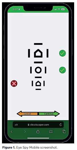
PURPOSE: This cross-sectional study compared best-corrected visual acuity obtained using Cloudscaper symbols, a novel optotype developed according to ETDRS specifications for children's virtual screening, with that obtained using LEA symbols.
METHODS: A total of 560 children aged 3-16 yr underwent visual acuity test with both Cloudscaper symbols and LS. The test application was standardized using the EyeSpy algorithm. Additionally, 147 participants were tested with the standard Snellen E paper chart. Paired t tests were performed to assess the clinical significance of logMAR visual acuity differences.
RESULTS: The mean logMAR visual acuity with LEA symbols was 0.12 (standard deviation [SD]=0.18; range, -0.10 to 0.80), while with Cloudscaper symbols it was 0.18 (SD=0.19; range, -0.10 to 0.80). The mean difference between Cloudscaper symbols and LEA symbols was 0.099 logMAR (approximately 0.5 optotypes; SD=0.08; range, 0.0-0.14; p<0.0001). Cloudscaper symbols slightly underestimated visual acuity compared to LEA symbols. Visual acuity measured by both methods was highly correlated (Spearman's r=0.74, p<0.0001). The mean visual acuity difference between Cloudscaper symbols and the Snellen E chart was 0.0045 (p=0.805; 95% confidence interval [95% CI]), whereas the difference between LEA symbols and Snellen E was 0.0883 (p<0.001; 95% CI).
CONCLUSIONS: Cloudscaper symbols provide a reliable tool for visual screening in children. Although they slightly underestimate visual acuity compared to LEA symbols – a finding also reported when comparing ETDRS letters with LEA symbols – Cloudscaper symbols show strong agreement with Snellen E chart measurements. This suggests that Cloudscaper symbols allow precise visual acuity assessment comparable to the gold standard.
Keywords: Vision screening; Vision tests; Visual acuity; Mobile applications; Eye health; Child health; Diagnostic techniques, Ophthalmological; Child; Preschool child; Adolescent
Arq. Bras. Oftalmol. 2025;88 (6 )
:1-5
| DOI: 10.5935/0004-2749.2025-0085
Abstract
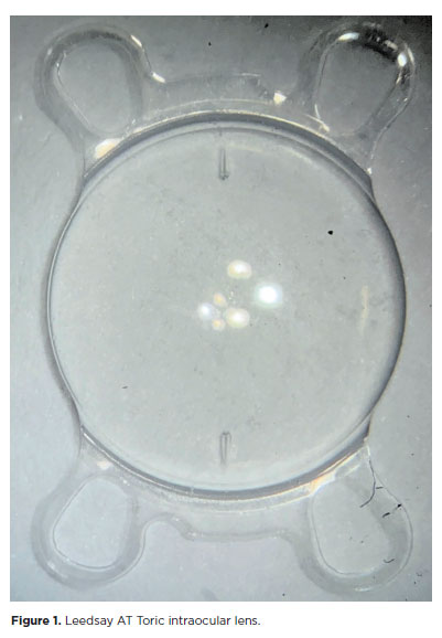
PURPOSE: The purpose of this study was to assess visual outcomes and patient satisfaction following cataract surgery involving the implantation of quad-loop intraocular lenses, including trifocal, bifocal, and toric variants.
METHODS: Information was obtained from both physical and electronic medical records of patients who underwent phacoemulsification cataract surgery with implantation of different intraocular lenses between January 1, 2022, and December 31, 2023. The study included individuals aged over 18 who received bilateral implantation of bifocal, trifocal, or monofocal toric intraocular lenses. Visual acuity was assessed at various postoperative time points using the logMAR scale. Quantitative variables were analyzed using mean and standard deviation.
RESULTS: A total of 92 eyes received premium intraocular lenses: 4 bifocal, 32 trifocal, 52 toric monofocal, and 4 trifocal toric lenses. The average preoperative corrected visual acuity was logMAR 0.478 ± 0.259. On the first postoperative day, the average uncorrected visual acuity was logMAR 0.301 ± 0.207. By day 30, 67.4% of eyes achieved uncorrected distance visual acuity of logMAR 0.2 or better. Patient satisfaction was high, with few reports of glare or halos.
CONCLUSION: Quad-loop intraocular lenses-including trifocal, bifocal, and toric models-demonstrated effective improvement in visual acuity and high levels of patient satisfaction. These lenses represent a suitable option for enhancing visual outcomes after cataract surgery. Additional studies with larger cohorts are recommended to confirm these results.
Keywords: Cataract extraction; Aberrometry/methods; Lenses, intraocular; Lens implantation, intraocular; Prosthesis design
Arq. Bras. Oftalmol. 2026;89 (2 )
:1-8
| DOI: 10.5935/0004-2749.2025-0175
Abstract
PURPOSE: Endophthalmitis is one of the most important adverse events after cataract surgery, as it can lead to total vision loss. This study aimed to describe the occurrence of endophthalmitis after phacoemulsification with intraocular lens implantation in patients treated in a community setting in Porto Velho, Rondônia, Brazil.
METHODS: This retrospective cohort study was conducted using a database of 649 medical records of patients who underwent surgery and were followed for three months. Poisson regression analysis was used to estimate relative risks and 95% confidence intervals (95% CIs).
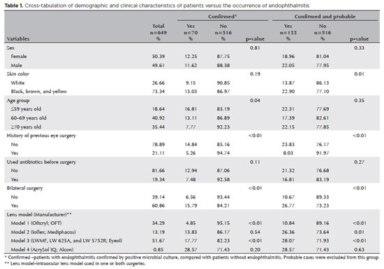
RESULTS: The incidence of confirmed endophthalmitis was 11.94% (95% CI, 9.50-14.76), while the incidence of confirmed and probable cases was 20.50% (95% CI, 17.52-23.73). For confirmed cases, bilateral surgery and the use of lens model 3 were identified as risk factors for endophthalmitis, whereas age over 70 yr and preoperative antibiotic use were protective factors. For confirmed and probable cases, brown and yellow skin color, bilateral surgery, and the use of lens model 3 were also identified as risk factors. Gram-negative bacteria were the predominant etiological agents, and corneal edema was the main clinical manifestation. The mean duration of treatment was eight days, and 27.12% of patients used antibiotics.
CONCLUSION: The incidence observed was substantially higher than that reported in the literature, with a predominance of Gram-negative agents and an association with bilateral surgeries and the Eyeol intraocular lens model. These findings reinforce the need for continuous epidemiological surveillance and the implementation of specific biosafety and infection control protocols during cataract surgery campaigns.
Keywords: Endophthalmitis; Disease outbreaks; Phacoemulsification; Lens implantation, intraocular; Lenses, intraocular; Cataract; Risk factors; Anti-bacterial agents
Arq. Bras. Oftalmol. 2025;88 (6 )
:1-8
| DOI: 10.5935/0004-2749.2024-0394
Abstract
The advantages and disadvantages of using perioperative subconjunctival steroid injections in dropless cataract surgery continue to be debated. A systematic review of PubMed, EMBASE, and the Cochrane Central database identified five studies—two randomized controlled trials and three non-randomized studies—encompassing 70,751 eyes. Among these, 12,319 eyes (17.4%) received subconjunctival steroid injections, while 58,432 eyes (82.6%) were managed with topical steroids. The Cochrane Collaboration’s RoB 2 tool was applied for bias assessments in randomized controlled trials, and heterogeneity was assessed using the I² statistics. No statistically significant differences were found between the two groups regarding macular edema (p=0.249), visual acuity (p=0.73), or laser flare count (p=0.45). Both subconjunctival injections and topical steroids demonstrated comparable efficacy and safety in controlling postoperative inflammation after cataract surgery. Additional research is warranted to validate these conclusions.
Keywords: Cataract extraction; Phacoemulsification; Lens implantation, intraocular; Postoperative care; Intravitreal injections; Anti-inflammatory agents, non-steroidal/administration & dosage; Glucocorticoids; Triamcinolone acetonide; Research design; Randomiz
Arq. Bras. Oftalmol. 2025;88 (2 )
:1-8
| DOI: 10.5935/0004-2749.2023-0268
Abstract
PURPOSE: This prospective, randomized, unmasked, clinical trial aimed to report the visual outcomes of cataract surgery on both eyes versus cataract surgery on one eye in Brazilian patients.
METHODS: This study included patients with bilateral cataracts and binocular visual acuity worse than or equal to 0.3 logarithm of the minimum angle of resolution. The patients were randomly assigned to undergo surgery on one (Control Group) or both eyes (one eye at a time; Intervention Group). Postoperatively, self-reported visual function using Catquest-9SF (primary outcome measure), binocular visual acuity, stereopsis, and ocular dominance (secondary outcome measures) were compared.
RESULTS: A total of 151 patients (77 and 148 eyes in the Control and Intervention Groups, respectively) completed the follow-up. Patients who underwent surgery on both eyes exhibited significantly better self-reported visual function (p=0.036) and stereopsis (p=0.026) than those who underwent surgery on one eye. Binocular visual acuity and ocular dominance did not affect the group comparisons.
CONCLUSIONS: Surgery on both eyes resulted in significantly better self-reported visual function and stereopsis than surgery on one eye.
Keywords: Cataract; Cataract extraction; Quality of life; Treatment outcome; Visual acuity; Binocular vision; Stereopsis
Arq. Bras. Oftalmol. 2026;89 (3 )
:1-4
| DOI: 10.5935/0004-2749.2025-0263
Abstract
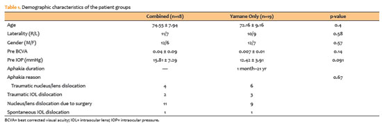
PURPOSE: To compare patients who underwent scleral fixation using the Yamane technique with and without simultaneous pars plana vitrectomy.
METHODS: A total of 37 patients were included in the study. Eighteen underwent simultaneous pars plana vitrectomy. The Yamane technique alone was performed only in patients with aphakia who had previously undergone pars plana vitrectomy for various reasons. Final lens position, best corrected visual acuity spherical equivalent, complication rates, and optical coherence tomography findings were recorded.
RESULTS: The duration of aphakia before intraocular lens implantation ranged from 1 month to 21 yr. Postoperative best corrected visual acuity improved in both groups, with no statistically significant difference (with pars plana vitrectomy: 0.42 ± 0.34; without pars plana vitrectomy: 0.32 ± 0.26; p=0.33). The spherical equivalent was also not significantly different between groups (with pars plana vitrectomy: 0.29 ± 1.08; without pars plana vitrectomy: 0.65 ± 2.23; p=0.53). There were no significant differences between the groups in complication rates, postoperative intraocular lens position or optical coherence tomography findings.
CONCLUSION: There was no difference in terms of safety or efficacy between the two approaches. Surgical decisions may be based on the surgeon’s experience and the patient’s systemic and ocular condition.
Keywords: Lens implantation, intraocular; Tomography, optical coherence; Vitrectomy; Intraocular lenses; Visual acuity; Aphakia; Yamane technique
Arq. Bras. Oftalmol. 2026;89 (4 )
:1-5
| DOI: 10.5935/0004-2749.2026-0010
Abstract
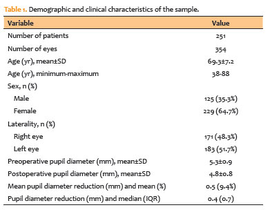
PURPOSE: To evaluate changes in scotopic pupil diameter before and after cataract surgery performed by phacoemulsification with intraocular lens implantation.
METHODS: This prospective longitudinal observational study included patients who underwent cataract surgery. Scotopic pupil diameter was measured preoperatively and 30-40 days postoperatively using an automated keratometer after a standardized dark-adaptation period under controlled ambient illumination. Each eye was considered an independent unit of observation. Because some participants contributed both eyes, intraindividual correlation was accounted for using a linear mixed-effects model with random patient intercepts. Time of assessment (preoperative versus postoperative), age, sex, and eye laterality were included as fixed effects.
RESULTS: A total of 354 eyes from 251 patients were analyzed. The mean patient age was 69.3±7.2 yr. Mean scotopic pupil diameter decreased from 5.3±0.9mm preoperatively to 4.8±0.8mm postoperatively, representing a mean reduction of 0.5mm (9.4%). In the linear mixed-effects model, cataract surgery was associated with a significant reduction in pupil diameter, with an adjusted mean difference of 0.45mm (95% confidence interval [95% CI], 0.39-0.51; p<0.001). Age (p=0.061), sex (p=0.920), and eye laterality (p=0.152) were not significantly associated with the magnitude of pupil diameter change.
CONCLUSION: Phacoemulsification with intraocular lens implantation was associated with a significant reduction in scotopic pupil diameter, independent of age, sex, and eye laterality. This finding should be considered during preoperative planning, particularly when selecting intraocular lenses whose optical performance depends on postoperative pupil size.
Keywords: Cataract; Pupil; Phacoemulsification; Lens implantation, intraocular; Lenses, intraocular; Pseudophakia
Arq. Bras. Oftalmol. 2025;88 (5 )
:1-7
| DOI: 10.5935/0004-2749.2024-0368
Abstract
PURPOSE: To compare endothelial corneal cell changes following cataract surgery performed by phacoemulsification with intraocular lens implantation, conducted by surgeons with varying levels of experience.
METHODS: Two hundred and eighty-three eyes diagnosed with cataract were included. Lens opacity was classified into three categories (I, II, and III). Surgeons were categorized into four experience levels (1, 2, 3, and 4), based on years of practice and lifetime surgeries performed. Corneal endothelial characteristics were assessed using non-contact specular microscopy, with measurements taken before surgery and 30-60 days post-surgery.
RESULTS: Pre- and postoperative endothelial analysis showed no significant differences between surgeon levels regarding visual acuity achieved, corneal thickness, and endothelial hexagonality. However, the central endothelial cell density index showed a significantly greater reduction among level 1 surgeons (p=0.026). Grade II cataracts exhibited significant variations in the central endothelial cell density (p=0.011) and average cell size, with level 1 surgeons showing the largest increases (p=0.024).
CONCLUSIONS: The analysis revealed significant differences in visual acuity and endothelial indices between surgeon experience levels, with less experienced surgeons showing greater variations and poorer performance. Clinical protocols should consider these data to establish safer training protocols.
Keywords: Cataract extraction; Phacoemulsification; Endothelium; corneal; Lens implantation, intraocular; Visual acuity; Internship and residency; Surgeons


 ABO is licensed under a Creative Commons Attribution-NonComercial 4.0 Internacional.


08-tab01.jpg)
06-tab01tb.jpg)
16-tab01tb.jpg)




06-fig01.jpg)
01-fig01.jpg)