Showing of 1 until 14 from 144 result(s)
Search for: Surgical wound dehiscence; Corneal transplantation; Risk factors
03-tab01.jpg)
Abstract
Objetivo: Investigar o impacto de diferentes tamanhos de incisões em córnea clara com meridiano íngreme para facoemulsificação com aberrações de mais alta ordem da córnea anterior.
Métodos: Foram retrospectivamente revisados os prontuários médicos de pacientes que se submeteram a cirurgias de catarata com microincisões coaxiais de 2,2 mm ou com incisões coaxiais pequenas de 2,75 mm. Foram apenas incluídos pacientes com astigmatismo preexistente da córnea anterior <2,00 dioptrias (D) e ≥0,50 D, e submetidos a incisões em córnea clara com meridiano íngreme. Os desfechos primários foram aberrações da córnea anterior da 3ª à 6ª ordem com uma pupila de 8 mm. O astigmatismo da córnea anterior e o tempo efetivo de facoemulsificação foram avaliados como desfechos secundários. Os desfechos pré-operatório e pós-operatório aos 3 meses também foram avaliados.
Resultados: O astigmatismo da córnea anterior diminuiu significativamente após ambos os procedimentos, mas não se encontrou nenhuma diferença significativa entre os dois procedimentos quanto ao astigmatismo da córnea anterior, induzido pela cirurgia (p=0,146). Embora as aberrações totais de mais alta ordem não se tenham alterado significativamente após ambos procedimentos, a comparação entre os grupos revelou uma diferença significativa nas aberrações totais de mais alta ordem, induzidas pela cirurgia (uma diminuição de 0,337 ± 1,156 μm na cirurgia de catarata por microincisão coaxial de 2,2 mm e um aumento de 0,106 ± 0,521 μm na cirurgia de catarata por incisão coaxial pequena de 2,75 mm; p=0,046). A aberração esférica diminuiu significativamente após cirurgia de catarata por microincisão coaxial de 2,2 mm (p=0,001), mas não se alterou significativamente após cirurgia de catarata por incisão coaxial pequena de 2,75 mm (p=0,564). A aberração de coma não mudou significativamente após qualquer dos procedimentos. O trifólio não se alterou significativamente após cirurgia de catarata por microincisão coaxial de 2,2 mm (p=0,361), mas aumentou significativamente após cirurgia de catarata por incisão coaxial pequena de 2,75 mm (p<0,001). Nenhuma diferença significativa se evidenciou quanto ao tempo efetivo de faco-emulsificação entre os dois procedimentos. Houve uma correlação positiva significativa entre o astigmatismo da córnea anterior, induzido pela cirurgia e a aberração de coma na cirurgia de catarata por incisão coaxial pequena de 2,75 mm (r=0,387, p=0,006). Não foi encontrada correlação significativa entre as alterações nas aberrações totais de mais alta ordem, induzidas pela cirurgia e o tempo efetivo de faco-emulsificação.
Conclusões: Nem a cirurgia de catarata por microincisão coaxial de 2,2 mm, nem aquela por incisão coaxial pequena de 2,75 mm degradaram significativamente as aberrações totais de mais alta ordem da córnea anterior. Porém, as alterações nas aberrações totais de mais alta ordem, induzidas pela cirurgia mostraram uma diferença significativa entre os dois procedimentos, com uma ligeira redução na cirurgia de catarata por microincisão coaxial de 2,2 mm e um pequeno aumento na cirurgia de catarata por incisão coaxial pequena de 2,75 mm. O tempo de facoemulsificação e a potência utilizada durante a cirurgia não tiveram impacto nas aberrações corneanas.
Keywords: Facoemuslificação; Astigmatismo; Cornea/cirurgia; Ferida cirúrgica; Resultado de tratamento
08-tab01tb.jpg)
Abstract
Objetivo: Estimar a epidemiologia do pterígio; sua correlação com sintomas de olho seco e com potenciais preditores sistêmicos e oculares.
Métodos: Estudo transversal, de base populacional, no qual foram realizadas visitas domiciliares aleatórias a 600 participantes, com 40 anos ou mais de idade, em Ribeirão Preto-SP (n=420) e Cassia dos Coqueiros-SP (n=180), Brasil. Uma entrevista estruturada com um questionário detalhado foi usada para coletar informações sobre demografia e possíveis fatores de risco. Em um segundo momento, participantes aleatórios com pterígio (n=63) ou não (n=110) foram avaliados quanto a alterações na superfície ocular.
Resultados: A frequência de pterígio em Ribeirão Preto foi de 21%; 15.7% entre as mulheres e 32.1% entre os homens (p=0,0002). Em Cássia dos Coqueiros, essa taxa foi de 19.4%; onde 17.3% eram mulheres e 25.5% eram homens (p=0,28). A média de idade naqueles afetados pelo pterígio foi superior à dos participantes sem pterígio, 65,6 ± 10,5 e 61,2 ± 12,0 anos, respectivamente (p=0,02). Houve uma correlação positiva entre o pterígio e história prévia de radioterapia e quimioterapia (p<0,0001 para ambos). Houve maior coloração de fluoresceína na córnea e maior coloração de lissamina verde na conjuntiva em olhos com pterígio (p=0,0003 e 0,0001, respectivamente).
Conclusão: Encontramos uma alta frequência de pterígio em duas populações adultas brasileiras, principalmente em homens e idosos. Danos na superfície ocular e história prévia de radioterapia e/ou quimioterapia foram associados ao pterígio.
Keywords: Pterígio/epidemiologia; Síndrome do olho seco; Prevalência; Fatores de risco
13-tab01.jpg)
Abstract
Objetivo: Avaliar o perfil clínico e epidemiológico dos transplantes de córnea realizados em um centro de referência oftalmológica de Recife no estado de Pernambuco, localizado no nordeste do Brasil.
Métodos: Esse estudo transversal coletou através de prontuários médicos dados clínicos e epidemiológicos de pacientes submetidos a ceratoplastia na Fundação Altino Ventura, de janeiro a dezembro de 2017.
Resultados: Um total de 356 procedimentos foram realizados em 327 pacientes dos quais 165 (50.5%) eram mulheres. A média de idade na cirurgia foi de 50.9 ± 22.6 anos (variação, 10 - 89 anos). A maioria dos pacientes (n=152 [46.5%]) era da capital e região metropolitana. A média de tempo de espera na fila para o transplante de córnea foi de 52.4 ± 58.9 dias (variação, 0 - 460 dias). As principais indicações de transplante foram ceratite infecciosa (n=88 [24.7%]), ceratocone (n=80 [22.5%]) e falência de transplante prévio (n=75 [21.1%]). Transplante penetrante foi a técnica mais realizada (n=213 [59.9%]) e foi mais comum em homens (n=132 [76.7%]), enquanto os transplantes lamelares posteriores (n=143 [41.1%]) foram mais realizados nas mulheres (p<0.001).
Conclusão: Ceratites infecciosas foram a causa mais comum de transplante, com prevalência similar em adultos economicamente ativos de ambos os sexos. Transplante penetrantes foram os prevalentes em homens e os transplantes lamelares em mulheres.
Keywords: Doença da córnea/epidemiologia; Transplante de córnea; Ceratoplastia penetrante; Brasil/epidemiologia
Abstract
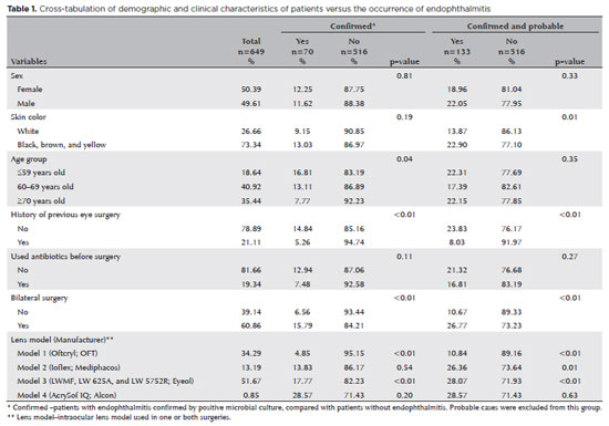
PURPOSE: Endophthalmitis is one of the most important adverse events after cataract surgery, as it can lead to total vision loss. This study aimed to describe the occurrence of endophthalmitis after phacoemulsification with intraocular lens implantation in patients treated in a community setting in Porto Velho, Rondônia, Brazil.
METHODS: This retrospective cohort study was conducted using a database of 649 medical records of patients who underwent surgery and were followed for three months. Poisson regression analysis was used to estimate relative risks and 95% confidence intervals (95% CIs).
RESULTS: The incidence of confirmed endophthalmitis was 11.94% (95% CI, 9.50-14.76), while the incidence of confirmed and probable cases was 20.50% (95% CI, 17.52-23.73). For confirmed cases, bilateral surgery and the use of lens model 3 were identified as risk factors for endophthalmitis, whereas age over 70 yr and preoperative antibiotic use were protective factors. For confirmed and probable cases, brown and yellow skin color, bilateral surgery, and the use of lens model 3 were also identified as risk factors. Gram-negative bacteria were the predominant etiological agents, and corneal edema was the main clinical manifestation. The mean duration of treatment was eight days, and 27.12% of patients used antibiotics.
CONCLUSION: The incidence observed was substantially higher than that reported in the literature, with a predominance of Gram-negative agents and an association with bilateral surgeries and the Eyeol intraocular lens model. These findings reinforce the need for continuous epidemiological surveillance and the implementation of specific biosafety and infection control protocols during cataract surgery campaigns.
Keywords: Endophthalmitis; Disease outbreaks; Phacoemulsification; Lens implantation, intraocular; Lenses, intraocular; Cataract; Risk factors; Anti-bacterial agents
Abstract
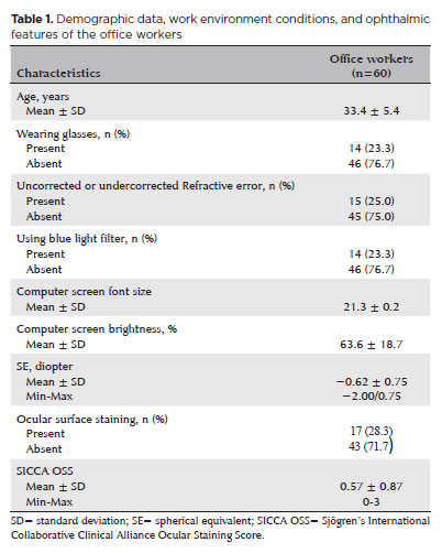
PURPOSE: To examine how ophthalmological features, screen exposure duration, and break habits among office employees affect ocular surface parameters.
METHODS: This single-center cross-sectional study involved two assessments on the same day: one before and one after a visual display terminal task. During the initial assessment, information on screen use was gathered, and refractive error, anterior segment examination, tear breakup time, and Schirmer test measurements were conducted. Participants tracked their screen usage and break durations throughout the day. At the end of the workday, tear breakup time and Schirmer I tests were repeated. Baseline and follow-up results were compared, and regression analysis was performed to identify factors linked to tear breakup time reduction.
RESULTS: The study enrolled 60 female office employees. Their mean screen time was 269.26 ± 70.21 min, with an average break duration of 151.93 ± 46.24 min. Tear breakup time at the second assessment (6.38 ± 2.70) was significantly lower than at baseline (8.62 ± 2.73) (p<0.001), whereas Schirmer test scores showed no significant change (p>0.05). Tear breakup time reduction was noted in 54 participants (90.0%), with a significant association between tear breakup time decrease percentage and screen exposure (p=0.001, r=0.463). Regression analysis showed that uncorrected or undercorrected refractive error was an independent risk factor for a ≥30% tear breakup time reduction, while taking more frequent short breaks (<15 min) acted as a protective factor.
CONCLUSIONS: Taking more frequent short breaks (<15 min) and correcting refractive errors help prevent intra-day tear breakup time decline during visual display terminal use. Structuring breaks to support tear film stability is advisable for occupations that require regular visual display terminal tasks.
Keywords: Tear film; Screen time; Tear breakup time; Office workers; Protective factors; Lacerations; Refractive errors; Risk factors.
06-tab01tb.jpg)
Abstract
MÉTODOS: Córneas humanas de treinamento disponibilizadas foram randomizadas em quatro grupos: Pachy-100 (profundidade de incisão = espessura corneana central - margem de segurança de 100 µm), Pachy-50 (margem de segurança de 50 µm), Pachy-0 (sem margem de segurança) e Pachy+50 (profundidade de incisão = espessura corneana central + 50 µm). Todas as lamelas foram dissecadas através um método padronizado e já publicado (Pachy-DSEK). As espessuras das lamelas (centro, zona de 3,0mm e zona de 6,0mm) foram medidas com tomografia de coerência óptica. A razão de espessura centro-periferia foi calculada aos 3,0 e 6,0 mm de diâmetro.
RESULTADOS: Perfuração endotelial ocorreu apenas no grupo Pachy+50 (n=3, 30%). A espessura central da lamela nos grupos Pachy-100, Pachy-50, Pachy-0 e Pachy+50 foi de 185 ± 42 µm, 122 ± 29 µm, 114 ± 29 µm, e 58 ± 31 µm, respectivamente (p<0,001). As razões C/P aos 3,0 e 6,0 mm foram de 0,97 ± 0,06 e 0,92 ± 0,14, respectivamente. Os parâmetros de características do doador não se correlacionaram com os resultados de espessura de lamela. A profundidade planejada de incisão se correlacionou com a maioria dos parâmetros de espessura de lamela (p<0,001). A espessura de lamela se correlacionou negativamente com a profundidade planejada da incisão (p<0.001, r=-0,580). O melhor resultado foi observado no grupo Pachy-0, em que 75% das lamelas mediram abaixo de 130 µm e não houve perfuração endotelial.
CONCLUSÃO: Através de um método padronizado de dissecção, a maioria das lamelas endoteliais apresentou uma configuração planar. O planejamento de profundidade de incisão igual à espessura corneana central resultou em alta porcentagem de lamelas ultrafinas sem ocorrência de perfuração.
Keywords: Transplante de córnea; Ceratoplastia lamelar; Endotélio corneano; Dissecção; Tomografia de coerência óptica
Abstract
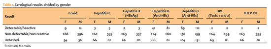
PURPOSE: To evaluate the impact of the COVID-19 pandemic and characterize the serological profile of discarded corneal donations in the coverage area of the Banco de Olhos de Londrina, through reverse transcription-polymerase chain reaction testing for COVID-19 and serological screening of cornea donors excluded because of positive test results.
METHODS: This observational retrospective study included 776 cornea donors who’s serological and reverse transcription-polymerase chain reaction test results were processed at the Hospital of Universidade Estadual de Londrina between May 2020 and 2022. The number of corneal donations and tissue utilization rates throughout the years of operation of the Banco de Olhos de Londrina were also analyzed.
RESULTS: The mean donor age was 53.14 years; 332 donors (43%) were female, and 444 (57%) were male. Positive results were identified in 15.76% of donors for hepatitis B core antibody antibodies, 0.65% for hepatitis B surface antigen, 1.03% for hepatitis C antibodies, and 0.52% for human immunodeficiency virus and human T-lymphotropic vírus. Positive reverse transcription-polymerase chain reaction results for SARS-CoV-2 were observed in 2.7% of cases. Older adults were 2.6 times more likely to test positive for SARS-CoV-2 (95% CI, 1.06-6.34) and 3.0 times more likely to test positive for hepatitis B core antibody (95% CI, 1.95-4.41) than younger individuals. A 75.2% reduction in corneal donations was observed in 2020 compared with 2019, accompanied by a 5% increase in tissue utilization, possibly associated with the effectiveness of donor screening during the pandemic.
CONCLUSION: The COVID-19 pandemic had a profound impact on the number of corneal transplants worldwide, in Brazil, and at the Banco de Olhos de Londrina because of the substantial decline in donations during this period. Hepatitis B was the leading cause of corneal tissue discard due to positive serology in both this study and previous reports, highlighting the importance of prevention programs and improved vaccination coverage. Strict legislation, comprehensive serological screening, and appropriate processing of donated tissue remain essential to eliminate potential sources of infection and ensure transplantation safety.
Keywords: Cornea; Corneal transplantation; COVID-19; Eye banks; Serology
Abstract
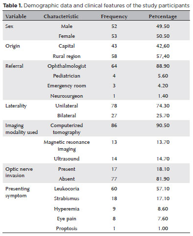
PURPOSE: Although Brazil has a high prevalence of retinoblastoma, there is a lack of epidemiological data on the disease. Thus, in this study, we aimed to evaluate the epidemiological profile of patients diagnosed with retinoblastoma in the ophthalmology department of a pediatric tertiary referral hospital in Ceara, Brazil.
METHODS: A descriptive and cross-sectional study was conducted by retrospectively analyzing the clinical and socioeconomic data from the medical records of pediatric patients followed-up at the hospital between 2007 and 2021. Retinoblastoma was diagnosed on the basis of a fundoscopic or histopathologic examination.
RESULTS: The data of 105 patients were included in the study, and the mean patient age at the time of diagnosis was 1.7 years. Most of the patients were women (50.5%) and hailed from rural areas (57.4%), which was associated with a higher tumor stage. Of the 150 patients, 57.1% initially presented with leukocoria. Ocular hyperemia was associated with more advanced stages of retinoblastoma (p=0.004). Bilateral involvement was observed in 25.7% of the patients and at a significantly younger age (p=0.009). The presence of retinal detachment, vascularized lesions, and vitreous seeds significantly increased the likelihood of requiring enucleation.
DISCUSSION: This study presents an epidemiological description of retinoblastoma in Brazil, which highlights the significance of early detection. Delayed diagnosis is associated with a poorer visual prognosis and higher mortality rate, particularly in patients with unilateral disease. Risk factors for a more severe disease were retinal detachment, vascularized lesions, and vitreous seeds. The correlation between histopathological features and clinical outcomes was limited.
CONCLUSION: Further studies are required to assess the influence of ocular hyperemia, fundoscopic assessment, and histopathologic findings on the prognosis of retinoblastoma. Moreover, it is critical to devise interventions to reduce the time-to-diagnosis in rural areas.
Keywords: Retinoblastoma; Retinal neoplasms; Epidemiology; Prevalence; Risk factors; Delayed diagnosis; Child
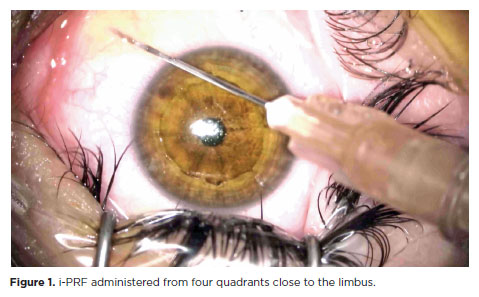
Abstract
PURPOSE: This study was conducted to investigate the effect of injectable platelet-rich fibrin on the recovery of compromised epithelium due to crosslinking treatment.
METHODS: In this comparative study, the epithelial closure rates and in vivo confocal biomicroscopy results of 26 patients with keratoconus who underwent subconjunctival injection of injectable platelet-rich fibrin near the limbus after epithelium-off corneal crosslinking treatment were compared with those of 25 patients who did not receive the injection of injectable platelet-rich fibrin.
RESULTS: The average time to epithelial defect closure in the injectable platelet-rich fibrin group was 2.76 ± 0.90 days compared to 3.56 ± 0.86 days in the non-injectable platelet-rich fibrin group (p=0.003). At the end of the 1st month, the mean subbasal nerve plexus density was 1.26 ± 1.61 nerves/mm2 in the injectable platelet-rich fibrin group, whereas it was 0.72 ± 0.89 nerves/mm2 in the non-injectable platelet-rich fibrin group (p=0.016). By the 3rd month, the density increased to 3.42 ± 1.13 nerves/mm2 in the injectable platelet-rich fibrin group and 2.36 ± 1.15 nerves/mm2 in the non-injectable platelet-rich fibrin group (p=0.002). Similarly, the anterior stromal keratocyte density at the end of the 1st month was 93.6 ± 33.5 cells/mm2 in the injectable platelet-rich fibrin group compared to 67.3 ± 26.4 cells/mm2 in the non-injectable platelet-rich fibrin group (p=0.001). By the end of the 3rd month, the density increased to 255.2 ± 45.7 cells/mm2 in the injectable platelet-rich fibrin group and 222.1 ± 43.6 cells/mm2 in the non-injectable platelet-rich fibrin group (p=0.011). In the non-injectable platelet-rich fibrin group, one patient developed a sterile infiltrate at the end of the 1st week, whereas no complications were observed in the injectable platelet-rich fibrin group.
CONCLUSION: Subconjunctival injectable platelet-rich fibrin application is an effective and safe method for corneal epithelial healing after crosslinking treatment.
Keywords: Keratoconus; Platelet-rich fibrin; Epithelium; corneal; Corneal crosslinking; Wound healing

Abstract
PURPOSE: Posterior capsule rupture is defined as an intraoperative posterior capsule tear resulting in vitreous loss. This study aimed to analyze the clinical characteristics, preoperative risk factors, intraoperative management strategies, and postoperative complications associated with posterior capsule rupture during phacoemulsification surgery.
METHODS: This was a retrospective observational cohort study of the medical records for 25,224 phacoemulsification surgeries performed at our tertiary eye care center between 2017 and 2022. We collected and collated the demographic characteristics and clinical findings of the patients in our cohort. Intraoperative management strategies and postoperative outcomes over a 1-year followup period were also recorded.
RESULTS: Posterior capsule rupture occurred in 351 eyes (351 patients), giving an overall posterior capsule rupture rate of 1.3%. The mean patient age was 68.6 ± 10.8 years. Pseudoexfoliation syndrome, mature cataracts, brown cataracts, and surgery performed by a resident were identified as risk factors for posterior capsule rupture (p<0.05 for each; the risk ratios were 2.70, 2.15, 2.44, 1.34, respectively). The most common intraoperative complications were dislocated lens fragments in the vitreous (8%) and iris damage (7.1%). The mean best-corrected visual acuity improved from 1.31 ± 0.84 (logMAR) postoperatively to 0.51 ± 0.56 at the end of the 1-year follow-up period (p<0.001). Corneal edema (55.6%) and elevated intraocular pressure (33.3%) were the most common early postoperative complications. Persistently elevated intraocular pressure (11.1%) and cystoid macular edema (5.1%) were the most common late postoperative complications.
CONCLUSION: Posterior capsule rupture is a common complication of phacoemulsification surgery that requires prolonged postoperative follow-up and a multidisciplinary approach. Despite the increased incidence of complications when rupture occurs, appropriate intraoperative and postoperative management can lead to satisfactory visual outcomes.
Keywords: Cataract extraction; Phacoemulsification; Posterior capsule rupture; Corneal edema; Risk factors; Postoperative complications; Intraoperative complications
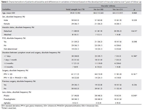
Abstract
PURPOSE: To clarify the postoperative incidence of macular edema in patients undergoing surgery to repair rhegmatogenous retinal detachment and identify the associated risk factors.
METHODS: In this prospective, observational study, 79 patients who underwent surgery to correct rhegmatogenous retinal detachment using pars plana vitrectomy with silicone oil injection were analyzed. Patients were followed up postoperatively at 7, 30, 90, 180, and 365 days. At each visit, optical coherence tomography was performed to assess the presence or absence of macular edema. were analyzed as possible risk factors for macular edema: age, sex, macular status (attached or detached), presence of vitreoretinal proliferation, history of previous intraocular surgery, reported time of symptoms suggestive of rhegmatogenous retinal detachment up to the date of surgery, and the surgical modality performed.
RESULTS: The 1-year macular edema prevalence rate was 26.6%. In the adjusted analysis, older patients had a higher risk of macular edema, and each 1-year increase in age increased the risk of macular edema by 6% (95% confidence interval = 1.00-1.12). The macular status, vitreoretinal proliferation, the surgical technique used, prior intraocular surgery, and the intraocular lens status were not identified as risk factors. However, the incidence of macular edema increased up to 180 days after surgery, peaking at 10.6%, and then decreased until 365 days after surgery.
CONCLUSION: Macular edema was a common complication after surgery to treat rhegmatogenous retinal detachment, with its incidence peaking between 30 and 180 days after surgery. Age was an important risk factor for macular edema in this cohort.
Keywords: Macular edema; Retinal detachment; Vitrectomy; Tomography, optical coherence; Incidence; Risk factors
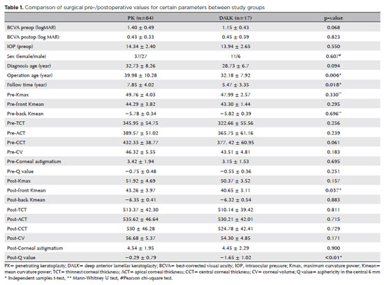
Abstract
PURPOSES: This study aims to assess and compare the postoperative visual and topographic outcomes, complications, and graft survival rates following deep anterior lamellar keratoplasty and penetrating keratoplasty in patients with macular corneal dystrophy.
METHODS: In this study we enrolled 59 patients (23 male; and 36 female) with macular corneal dystrophy comprising 81 eyes. Out of these, 64 eyes underwent penetrating keratoplasty, while 17 eyes underwent deep anterior lamellar keratoplasty. The two groups were analyzed and compared based on best-corrected visual acuity, corneal tomography parameters, pachymetry, complication rates, and graft survival rates.
RESULTS: After 12 months, 70.6% of the patients who underwent deep anterior lamellar keratoplasty (DALK) and 75% of those who had penetrating keratoplasty (PK) achieved a best-corrected visual acuity of 20/40 or better (p=0.712). Following surgery, DALK group showed lower front Kmean (p=0.037), and Q values (p<0.01) compared to the PK group. Postoperative interface opacity was observed in seven eyes (41.2%) in the DALK group. Other topography values and other complications (graft rejection, graft failure, cataract, glaucoma, microbial keratitis, optic atrophy) did not show significant differences between the two groups. The need for regrafting was 9.4% and 11.8% in the PK and DALK groups, respectively (p=0.769). Graft survival rates were 87.5% and 88.2% for PK and DALK; respectively (p=0.88 by Log-rank test).
CONCLUSION: Both PK and DALK are equally effective in treating macular corneal dystrophy, showing similar visual, topographic, and survival outcomes. Although interface opacity occurs more frequently after DALK the visual results were comparable in both groups. Therefore, DALK emerges as a viable surgical choice for patients with macular corneal dystrophy without Descemet membrane involvement is absent.
Keywords: Macular corneal dystrophy; Corneal dystrophies; Hereditary; Keratoplasty; Penetrating; Corneal transplantation
12-fig01.jpg)
Abstract
A retinopatia lúpica é uma manifestação clínica do lúpus eritematoso sistêmico no sistema visual. Geralmente assintomática, porém pode ser uma condição ameaçadora à visão. Está intimamente associada à atividade inflamatória do lúpus eritematoso sistêmico e ao aumento da mortalidade. A retinopatia lúpica tem diversas apresentações clínicas, como a microangiopatia lúpica, oclusão vascular, vasculite, retinopatia hipertensiva associada à nefrite lúpica e retinopatia autoimune. A prevalência e os fatores associados à retinopatia lúpica estão bem definidos em algumas partes do mundo. No entanto, esses dados são pouco conhecidos na América Latina, incluindo o Brasil. Como a retinopatia lúpica é geralmente assintomática, sem a fundoscopia de rotina, provavelmente esta é subestimada. O objetivo desta revisão é discutir a epidemiologia e fatores de risco para retinopatia lúpica.
Keywords: Lúpus eritematoso sistêmico/epidemiologia; Doenças retinianas; Fatores de risco
06-fig01.jpg)
Abstract
O presente relato de caso identificou a maculopatia média aguda paracentral como a causa de baixa de acuidade visual severa e irreversível após cirurgia de catarata. Existem fatores de risco bem estabelecidos para o desenvolvimento da maculopatia média aguda paracentral que devem ser conhecidos pelos cirurgiões de catarata. Nesse contexto cirúrgico, precauções extras no tocante a procedimentos anestésicos, pressão intraocular e alguns outros aspectos da cirurgia devem ser consideradas. A maculopatia média aguda paracentral é descrita como um sinal clínico observado no exame de tomografia de coerência óptica por domínio espectral e se trata, provavelmente, da evidência de um evento isquêmico no tecido vascular retiniano. Esse diagnóstico deve ser cogitado nos casos de perda de acuidade visual súbita no pós-operatório imediato associada com exame fundoscópico normal, como evidenciado no caso apresentado.
Keywords: Tomografia, coerência óptica; Procedimentos cirúrgicos oftalmológicos; Complicações pós-operatórias; Fatores de risco; Catarata; Extração de catarata; Baixa visão; Saúde oc
 ABO is licensed under a Creative Commons Attribution-NonComercial 4.0 Internacional.
About
Issues
Editorial Board
Submission
Arquivos Brasileiros de Oftalmologia
Official publication of Brazilian Council of Ophthalmology - Conselho Brasileiro de Oftalmologia (CBO)
Rua Casa do Ator, 1.117 - 2nd floor - Zip Code: 04546-004
São Paulo - SP, Brazil
TEL: +55 11 3266-4000
E-mail: [email protected]