Arq. Bras. Oftalmol. 2021;84 (3 )
:214-219
| DOI: 10.5935/0004-2749.20210029
Abstract
OBJETIVO: Avaliar a influência da dinâmica pupilar na curva de desfoco de olhos implantados com lente intraoculares multifocais difrativas.
MÉTODOS: Estudo prospectivo e randomizado realizado na Faculdade de Medicina de Ribeirão Preto - Universidade de São Paulo - Departamento de Oftalmologia. Trinta e oito pacientes foram aleatoriamente designados para receber bilateralmente lentes intraoculares SN6AD1 (n=20) (mfIOL) ou SN60WF (n=18) (aIOL). Além da acuidade visual para longe e perto, corrigida e não corrigida, e curva de desfoco, foi ainda realizada pupilometria dinâmica. A área sob a curva de desfoco foi calculada usando um modelo polinomial empírico.
RESULTADOS: Um total de 16 e 17 pacientes (n=32 e 34 olhos) completaram 1 ano de seguimento nos grupos mfIOL e aIOL, respectivamente. Não houve diferenças significativas entre grupos para as acuidades visuais seja para longe ou perto. As curvas de desfoco do grupo mfIOL mostraram um pico duplo; enquanto o SN60WF mostrou apenas um pico, típico para uma lente intraoculares monofocal. A média da área sob a curva de desfoco do grupo aIOL foi (4,66 ± 1,51 logMAR.dp), e essa é estatisticamente significante diferente da métrica do grupo mfIOL (1,99 ± 1,31 logMAR.dp). A pupila na contração máxima após a exposição a um flash de 30 cd/m2 por 1 segundo foi significativamente correlacionada com uma melhor área de foco no grupo mfIOL (r=0,54; p=0,0017), essa relação não foi observada para o grupo aIOL.
CONCLUSÃO: Estes dados indicam que quanto menor a pupila durante contração, melhor é a área sob a curva de desfoco e, portanto, o desempenho visual dos olhos implantados com essa mfIOL. Esta correlação não foi encontrada para lentes intraoculares monofocais.
Keywords: Lentes intraoculares multifocais; Pupila/fisiologia, Catarata; Facoemulsificacão
Arq. Bras. Oftalmol. 2023;86 (2 )
:113-120
| DOI: 10.5935/0004-2749.20230022
Abstract
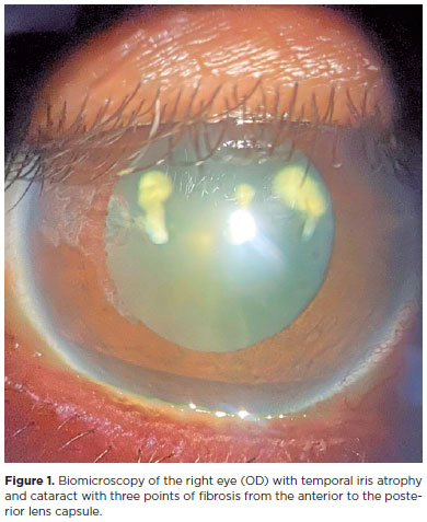
Objetivos: Avaliar a estabilidade e eficácia da técnica double-flanged com sutura de 5-0 polipropileno para fixação de cataratas subluxadas aos 18 meses e as possíveis complicações desta nova técnica.
Métodos: Esta técnica utiliza um monofilamento de polipropileno 5-0 para criar dois flanges com um termocautério para fixar um Segmento de Tensão Capsular na esclera a fim de estabilizar o saco capsular subluxado. Esta técnica foi implementada em 17 olhos que necessitavam do implante de lente intraocular em casos de diálise zonular devido a trauma, síndrome de Marfan, microesferofacia, subluxação idiopática ou pós-facoemulsificação que provocou subulxação do saco capsular intraoperatória.
Resultados: O seguimento dos pacientes foi de 18 meses. A acuidade visual corrigida melhorou significativamente de 0,85 para 0,39 (logMAR), enquanto os erros de refração esféricos e cilíndricos e a pressão intraocular permaneceram estáveis. Nenhuma fotodegradação de sutura ou pseudofacodonese foi encontrada.
Conclusão: A técnica double-flanged para fixação transescleral de saco capsular com sutura de 5-0 polipropileno mostrou resultados de estabilidade de longo prazo para o complexo lente/saco capsular. Então, aparenta ser uma opção segura para cirurgia de catarata, sem necessidade pontos, em olhos com fraqueza zonular ou diálise
Keywords: Catarata; Facoemulsificação; Lente intraocular; Técnica de sutura; Acuidade visual
Arq. Bras. Oftalmol. 2022;85 (4 )
:359-363
| DOI: 10.5935/0004-2749.20220046
Abstract
Objetivo: Investigar os resultados pós-operatórios e avaliar os preditores de sucesso da facoemulsificação combinada à goniotomia com o Kahook Dual Blade para o tratamento da catarata e do glaucoma em olhos com glaucoma primário de ângulo aberto.
Métodos: Série de casos retrospectivos, não comparativos e intervencionistas, em que todos os pacientes com glaucoma primário de ângulo aberto submetidos ao procedimento de facoemulsificação combinada à goniotomia com o Kahook Dual Blade entre junho de 2018 e abril de 2019 foram inscritos. Todos os participantes tiveram um acompanhamento mínimo de 6 meses. Foram registrados os valores de pressão intraocular pré e pós-operatória (em 1, 3 e 6 meses), número de medicamentos antiglaucomatosos, melhor acuidade visual corrigida, complicações cirúrgicas e quaisquer eventos ou procedimentos subsequentes relacionados. A análise de regressão logística foi usada para investigar a associação entre diferentes variáveis e resultados cirúrgicos.
Resultados: Um total de 57 olhos de 47 pacientes foram incluídos (média de idade, 70,5 ± 7 anos). A pressão intraocular média reduziu de 15,5 ± 4,2 mmHg para 12,2 ± 2,4 mmHg na última visita de acompanhamento (p<0,001). O número médio de medicamentos antiglaucomatosos diminuiu significativamente de 1,9 ± 1,0 para 0,6 ± 1,0 durante o mesmo período (p<0,001). Com base no critério predefinido (redução da pressão intraocular ≥20% e/ou redução de ≥1 medicamento), a taxa de sucesso em 6 meses foi de 86%. Um valor de pressão intraocular pré-operatório mais alto (OR= 2,01; p=0,016) e maior porcentagem de redução da pressão intraocular inicial
(30 dias) (OR= 1,02; p=0,033) foram significativamente associados ao sucesso cirúrgico.
Conclusão: Nossos resultados sugerem que o procedimento de facoemulsificação combinada à goniotomia com o Kahook Dual Blade é uma alternativa eficaz e segura para o manejo da catarata em olhos com glaucoma primário de ângulo aberto, impactando positivamente no controle da pressão intraocular e no número de medicamentos. Olhos com pressão intraocular basal mais alta e resposta inicial mais pronunciada ao procedimento parecem apresentar melhores resultados em 6 meses. Mais estudos são necessários para avaliar a eficácia em longo prazo e o perfil de segurança.
Keywords: Glaucoma; Glaucoma de ângulo aberto; Catarata; Facoemulsificação; Pressão intraocular; Goniotomia
Arq. Bras. Oftalmol. 2022;85 (3 )
:249-254
| DOI: 10.5935/0004-2749.20220036
Abstract
Objetivo: Criar modelos, em catarata pediátrica, para estimar valores futuros de ceratometria e comprimento axial, com base na ceratometria e no comprimento axial medidos na cirurgia, para previsão do poder da lente intraocular para emetropia em idades futuras.
Métodos: Olhos com catarata bilateral, ceratometria e comprimento axial medidos na cirurgia e pelo menos um exame pós-operatório com medidas de ceratometria e comprimento axial foram considerados para este estudo. Os modelos para estimar futuras ceratometrias e comprimentos axiais foram criados considerando (1) ceratometria e comprimento axial medidos na cirurgia, (2) a inclinação média da regressão logarítmica da ceratometria e comprimento axial criada para cada olho e (3) a idade na cirurgia. A lente intraocular para emetropia em idades futuras pode ser estimada usando esses valores em fórmulas de terceira geração. Os erros de estimativa da ceratometria, comprimento axial e poder da lente intraocular, usando os modelos, também foram calculados.
Resultados: 57 olhos de 29 pacientes preencheram os critérios de inclusão. A idade média na cirurgia e acompanhamento foram de 36,96 ± 32,04 meses e 2,39 ± 1,46 anos, respectivamente. A inclinação média da regressão logarítmica criada para cada olho foi de -3.286 para ceratometria e + 3.189 para o comprimento axial. Os erros médios de estimativa absoluta para ceratometria e comprimento axial foram respectivamente: 0,61 ± 0,54 D e 0,49 ± 0,55 mm, e para o poder da lente intraocular usando as fórmulas SRK-T, Hoffer-Q e Holladay I foram: 2,04 ± 1,73 D, 2,49 ± 2,10 D e 2,26 ± 1,87 D, respectivamente.
Conclusões: Os modelos apresentados podem ser utilizados para estimar o poder da lente intraocular que levaria a emetropia em idades futuras e orientar a escolha do poder da lente intraocular a ser implantada na catarata pediátrica.
Keywords: Catarata; Biometria/métodos; Emetropia; Comprimento axial do olho; Lentes intraoculares; Criança
Arq. Bras. Oftalmol. 2026;89 (1 )
:1-6
| DOI: 10.5935/0004-2749.2025-0052
Abstract
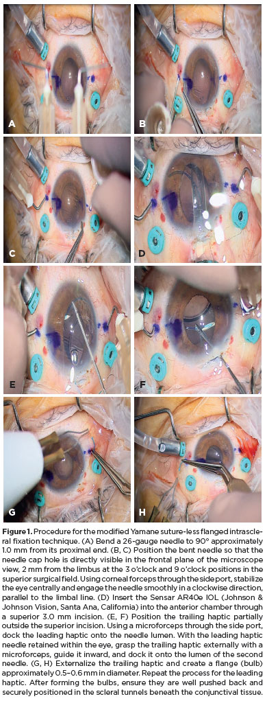
PURPOSE: To evaluate whether two simplified modifications of flanged intrascleral fixation techniques (Yamane and Canabrava) provide comparable refractive outcomes and complication rates while reducing surgical complexity in trocar-assisted vitrectomy.
METHODS: This retrospective observational study included 88 patients who underwent flanged fixation surgery with vitrectomy. In the modified Yamane technique, a single-path sclerotomy with bilateral symmetry was performed instead of an angled sclerotomy. In the modified Canabrava technique, the intraocular lens was inserted first, followed by the creation of a circular polypropylene loop with 2-mm flange spacing. Postoperative refractive parameters, including intraocular lens astigmatism, and complications such as intraocular lens iris capture were analyzed.
RESULTS: Of the 88 patients, 70 underwent the modified Yamane technique, and 18 underwent the modified Canabrava technique. No significant differences were observed between the two techniques regarding refractive outcomes or postoperative complications, except for surgical duration, which was significantly shorter (p<0.001) in one technique. Mean intraocular lens astigmatism was −0.675 D for Yamane and −0.666 D for Canabrava.
CONCLUSION: Optimizing needle engagement for symmetry in the Yamane technique and narrowing flange spacing while ensuring a circular polypropylene configuration in the Canabrava technique may reduce surgical complexity and improve postoperative outcomes.
Keywords: Polypropylenes; Yamane technique; Vitrectomy; Astigmatism; Lenses, intraocular; Postoperative complications; Suture techniques; Iris.
Arq. Bras. Oftalmol. 2026;89 (2 )
:1-8
| DOI: 10.5935/0004-2749.2025-0175
Abstract
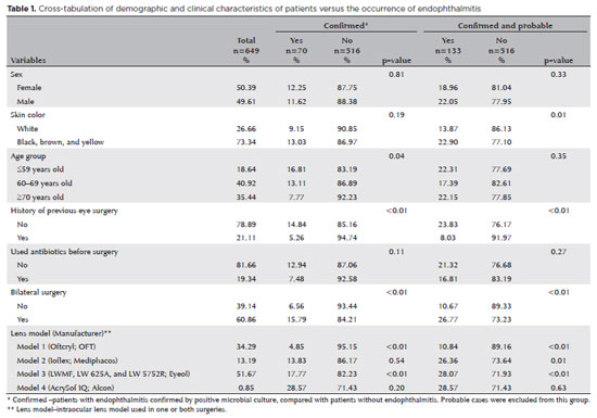
PURPOSE: Endophthalmitis is one of the most important adverse events after cataract surgery, as it can lead to total vision loss. This study aimed to describe the occurrence of endophthalmitis after phacoemulsification with intraocular lens implantation in patients treated in a community setting in Porto Velho, Rondônia, Brazil.
METHODS: This retrospective cohort study was conducted using a database of 649 medical records of patients who underwent surgery and were followed for three months. Poisson regression analysis was used to estimate relative risks and 95% confidence intervals (95% CIs).
RESULTS: The incidence of confirmed endophthalmitis was 11.94% (95% CI, 9.50-14.76), while the incidence of confirmed and probable cases was 20.50% (95% CI, 17.52-23.73). For confirmed cases, bilateral surgery and the use of lens model 3 were identified as risk factors for endophthalmitis, whereas age over 70 yr and preoperative antibiotic use were protective factors. For confirmed and probable cases, brown and yellow skin color, bilateral surgery, and the use of lens model 3 were also identified as risk factors. Gram-negative bacteria were the predominant etiological agents, and corneal edema was the main clinical manifestation. The mean duration of treatment was eight days, and 27.12% of patients used antibiotics.
CONCLUSION: The incidence observed was substantially higher than that reported in the literature, with a predominance of Gram-negative agents and an association with bilateral surgeries and the Eyeol intraocular lens model. These findings reinforce the need for continuous epidemiological surveillance and the implementation of specific biosafety and infection control protocols during cataract surgery campaigns.
Keywords: Endophthalmitis; Disease outbreaks; Phacoemulsification; Lens implantation, intraocular; Lenses, intraocular; Cataract; Risk factors; Anti-bacterial agents
Arq. Bras. Oftalmol. 2026;89 (1 )
:1-9
| DOI: 10.5935/0004-2749.2025-0097
Abstract
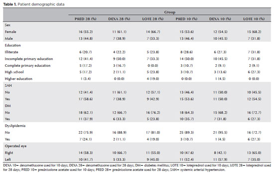
PURPOSE: To evaluate the efficacy of different corticosteroid eye drop formulations (prednisolone acetate 1.0%, dexamethasone 1.0%, and loteprednol etabonate 0.5%) administered for different treatment durations (10 vs. 28 days) in controlling postoperative inflammation following uncomplicated cataract surgery.
METHODS: This randomized, masked clinical trial was conducted at the Instituto Cearense de Oftalmologia. Eligible participants were aged ≥50 yr and scheduled for routine cataract surgery. Exclusion criteria included preexisting ocular disease (elevated intraocular pressure, retinopathy, maculopathy, or uveitis) or concurrent medication use that could confound results. Patients were randomized to receive prednisolone acetate (1.0%), dexamethasone (1.0%), or loteprednol etabonate (0.5%) four times daily for 28 days (with tapering) or for 10 days. Medication bottles, prescriptions, and examiners were masked. Postoperative assessments included ocular symptoms, visual acuity, intraocular pressure, anterior chamber cell count and flare, pachymetry, endothelial cell density, and macular thickness over a 30-day follow-up.
RESULTS: A total of 140 eyes from 140 patients were analyzed (29 prednisolone acetate 1.0%, 18 dexamethasone 1.0%, and 21 loteprednol etabonate 0.5% for 28 days; 28 prednisolone acetate 1.0%, 22 dexamethasone 1.0%, and 22 loteprednol etabonate 0.5% for 10 days). No significant differences were found among the six groups during follow-up. However, eyes treated with dexamethasone (1.0%) showed greater intraocular pressure fluctuations, particularly on Days 7 and 30, and a higher incidence of rebound inflammation in the 28-day regimen. Structural cystoid macular edema without visual impact was observed in 5.9% of eyes in the 28-day groups and 14.2% of eyes in the 10-day groups, as detected by optical coherence tomography at 30 days.
CONCLUSION: Equivalent postoperative inflammation control can be achieved using different corticosteroid eye drop formulations at varying treatment durations following cataract surgery. Brazilian Registry of Clinical Trials (ReBEC): RBR-2frpntv
Keywords: Adrenal cortex hormones; Cataract; Cystoid macular edema; Corticosteroids; Inflammation; Loteprednol etabonate; Ophthalmic solutions; Postoperative period; Intraocular pressure; Visual acuity
Arq. Bras. Oftalmol. 2025;88 (6 )
:1-8
| DOI: 10.5935/0004-2749.2024-0394
Abstract
The advantages and disadvantages of using perioperative subconjunctival steroid injections in dropless cataract surgery continue to be debated. A systematic review of PubMed, EMBASE, and the Cochrane Central database identified five studies—two randomized controlled trials and three non-randomized studies—encompassing 70,751 eyes. Among these, 12,319 eyes (17.4%) received subconjunctival steroid injections, while 58,432 eyes (82.6%) were managed with topical steroids. The Cochrane Collaboration’s RoB 2 tool was applied for bias assessments in randomized controlled trials, and heterogeneity was assessed using the I² statistics. No statistically significant differences were found between the two groups regarding macular edema (p=0.249), visual acuity (p=0.73), or laser flare count (p=0.45). Both subconjunctival injections and topical steroids demonstrated comparable efficacy and safety in controlling postoperative inflammation after cataract surgery. Additional research is warranted to validate these conclusions.
Keywords: Cataract extraction; Phacoemulsification; Lens implantation, intraocular; Postoperative care; Intravitreal injections; Anti-inflammatory agents, non-steroidal/administration & dosage; Glucocorticoids; Triamcinolone acetonide; Research design; Randomiz
Arq. Bras. Oftalmol. 2025;88 (2 )
:1-7
| DOI: 10.5935/0004-2749.2023-0215
Abstract
PURPOSE: To compare the refractive prediction error of Hill-radial basis function 3.0 with those of 3 conventional formulas and 11 combination methods in eyes with short axial lengths.
METHODS: The refractive prediction error was calculated using 4 formulas (Hoffer Q, SRK-T, Haigis, and Hill-RBF) and 11 combination methods (average of two or more methods). The absolute error was determined, and the proportion of eyes within 0.25-diopter (D) increments of absolute error was analyzed. Furthermore, the intraclass correlation coefficients of each method were computed to evaluate the agreement between target refractive error and postoperative spherical equivalent.
RESULTS: This study included 87 eyes. Based on the refractive prediction error findings, Hoffer Q formula exhibited the highest myopic errors, followed by SRK-T, Hill-RBF, and Haigis. Among all the methods, the Haigis and Hill-RBF combination yielded a mean refractive prediction error closest to zero. The SRK-T and Hill-RBF combination showed the lowest mean absolute error, whereas the Hoffer Q, SRK-T, and Haigis combination had the lowest median absolute error. Hill-radial basis function exhibited the highest intraclass correlation coefficient, whereas SRK-T showed the lowest. Haigis and Hill-RBF, as well as the combination of both, demonstrated the lowest proportion of refractive surprises (absolute error >1.00 D). Among the individual formulas, Hill-RBF had the highest success rate (absolute error ≤0.50 D). Moreover, among all the methods, the SRK-T and Hill-RBF combination exhibited the highest success rate.
CONCLUSIONS: Hill-radial basis function showed accuracy comparable to or surpassing that of conventional formulas in eyes with short axial lengths. The use and integration of various formulas in cataract surgery for eyes with short axial lengths may help reduce the incidence of refractive surprises.
Keywords: Cataract; Lenses, intraocular; Axial length, eye; Refractive errors; Artificial intelligence
Arq. Bras. Oftalmol. 2025;88 (5 )
:1-7
| DOI: 10.5935/0004-2749.2024-0217
Abstract
PURPOSE: This study aimed to evaluate the influence of intrastromal corneal ring segment implants on the intraocular pressure measurements using Goldmann applanation tonometry, rebound tonometry, and noncontact tonometry in keratoconic corneas and analyze the intertonometer agreement.
METHODS: We enrolled 74 eyes in this observational and prospective study. Each participant had a complete eye examination, corneal analysis with Scheimpflug Tomography (Pentacam®), and intraocular pressure evaluation with Goldmann applanation tonometry, rebound tonometry, and noncontact tonometry, before and after intrastromal corneal ring segment implantation (on postoperative days 1, 7, 45, and 90). Intertonometer agreement was assessed using Bland-Altman analysis.
RESULTS: The mean age was 29.9 ± 10.2 years, and 47 (63.5%) eyes had keratoconus grade II. Intraocular pressures were higher for noncontact tonometry preoperatively and on 90 postoperative day (mean ± SD: 12.4 ± and 12.1 ± 2.2 mmHg, respectively), followed by Goldmann applanation tonometry (11.1 ± 3.0 and 11.2 ± 2.7 mmHg, respectively), and were lower for rebound tonometry (9.7 ± and 9.4 ± 3.2 mmHg, respectively). The variation from the Goldmann tonometry on 7 postoperative day to the baseline (p=0.022) and that of noncontact tonometry on 90 postoperative day to the baseline (p=0.021) were statistically significant. The rebound tonometry underestimated intraocular pressure when compared with the Goldmann applanation tonometry by a mean of 1.47 ± 5.19 mmHg. Noncontact tonometry, when compared with Goldmann applanation tonometry, overesti-mated intraocular pressure by a mean of 1.23 ± 4.15 mmHg.
CONCLUSION: Despite statistically significant differences between some postoperative periods, the intraocular pressure measurement differences may not be clinically relevant.
Keywords: Keratoconus; Intraocular pressure; Cornea; Corneal stroma; Postoperative period; Tonometry ocular; Prostheses and implants
Arq. Bras. Oftalmol. 2026;89 (4 )
:1-5
| DOI: 10.5935/0004-2749.2026-0010
Abstract
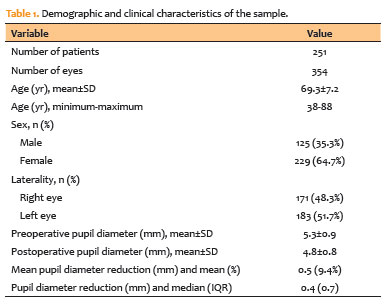
PURPOSE: To evaluate changes in scotopic pupil diameter before and after cataract surgery performed by phacoemulsification with intraocular lens implantation.
METHODS: This prospective longitudinal observational study included patients who underwent cataract surgery. Scotopic pupil diameter was measured preoperatively and 30-40 days postoperatively using an automated keratometer after a standardized dark-adaptation period under controlled ambient illumination. Each eye was considered an independent unit of observation. Because some participants contributed both eyes, intraindividual correlation was accounted for using a linear mixed-effects model with random patient intercepts. Time of assessment (preoperative versus postoperative), age, sex, and eye laterality were included as fixed effects.
RESULTS: A total of 354 eyes from 251 patients were analyzed. The mean patient age was 69.3±7.2 yr. Mean scotopic pupil diameter decreased from 5.3±0.9mm preoperatively to 4.8±0.8mm postoperatively, representing a mean reduction of 0.5mm (9.4%). In the linear mixed-effects model, cataract surgery was associated with a significant reduction in pupil diameter, with an adjusted mean difference of 0.45mm (95% confidence interval [95% CI], 0.39-0.51; p<0.001). Age (p=0.061), sex (p=0.920), and eye laterality (p=0.152) were not significantly associated with the magnitude of pupil diameter change.
CONCLUSION: Phacoemulsification with intraocular lens implantation was associated with a significant reduction in scotopic pupil diameter, independent of age, sex, and eye laterality. This finding should be considered during preoperative planning, particularly when selecting intraocular lenses whose optical performance depends on postoperative pupil size.
Keywords: Cataract; Pupil; Phacoemulsification; Lens implantation, intraocular; Lenses, intraocular; Pseudophakia
Arq. Bras. Oftalmol. 2025;88 (5 )
:1-8
| DOI: 10.5935/0004-2749.2024-0328
Abstract
PURPOSE: Posterior capsule rupture is defined as an intraoperative posterior capsule tear resulting in vitreous loss. This study aimed to analyze the clinical characteristics, preoperative risk factors, intraoperative management strategies, and postoperative complications associated with posterior capsule rupture during phacoemulsification surgery.
METHODS: This was a retrospective observational cohort study of the medical records for 25,224 phacoemulsification surgeries performed at our tertiary eye care center between 2017 and 2022. We collected and collated the demographic characteristics and clinical findings of the patients in our cohort. Intraoperative management strategies and postoperative outcomes over a 1-year followup period were also recorded.
RESULTS: Posterior capsule rupture occurred in 351 eyes (351 patients), giving an overall posterior capsule rupture rate of 1.3%. The mean patient age was 68.6 ± 10.8 years. Pseudoexfoliation syndrome, mature cataracts, brown cataracts, and surgery performed by a resident were identified as risk factors for posterior capsule rupture (p<0.05 for each; the risk ratios were 2.70, 2.15, 2.44, 1.34, respectively). The most common intraoperative complications were dislocated lens fragments in the vitreous (8%) and iris damage (7.1%). The mean best-corrected visual acuity improved from 1.31 ± 0.84 (logMAR) postoperatively to 0.51 ± 0.56 at the end of the 1-year follow-up period (p<0.001). Corneal edema (55.6%) and elevated intraocular pressure (33.3%) were the most common early postoperative complications. Persistently elevated intraocular pressure (11.1%) and cystoid macular edema (5.1%) were the most common late postoperative complications.
CONCLUSION: Posterior capsule rupture is a common complication of phacoemulsification surgery that requires prolonged postoperative follow-up and a multidisciplinary approach. Despite the increased incidence of complications when rupture occurs, appropriate intraoperative and postoperative management can lead to satisfactory visual outcomes.
Keywords: Cataract extraction; Phacoemulsification; Posterior capsule rupture; Corneal edema; Risk factors; Postoperative complications; Intraoperative complications
 ABO is licensed under a Creative Commons Attribution-NonComercial 4.0 Internacional.
03-fig01.jpg)
08-tab01.jpg)
03-fig01.jpg)
07-tab01tb.jpg)


10-fig01.jpg)
01-fig01.jpg)